photo free
Home Images Vectors Videos

Stock image Immune System Response page 2

stock image Immune response to coronavirus infection: coronaviruses marked bei antibodies are engulfed and destroyed by a macrophage immune cell

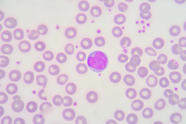
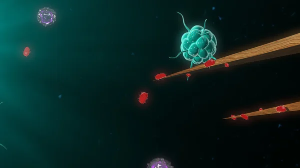
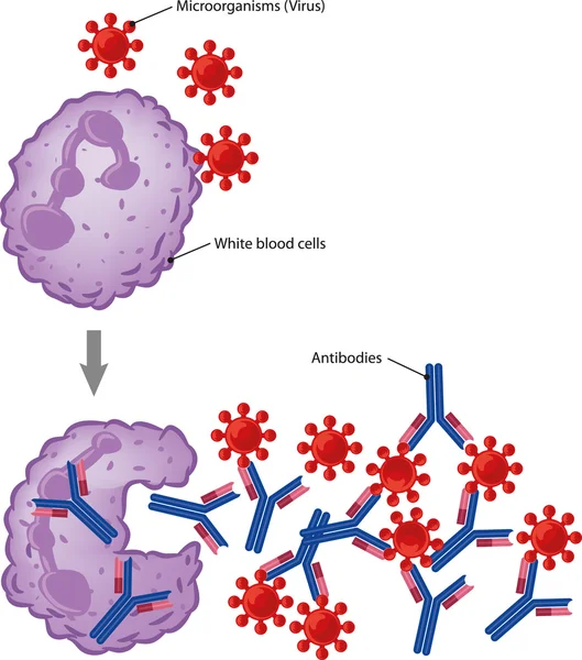
Immune Response To Coronavirus Infection: Coronaviruses Marked Bei Antibodies Are Engulfed And Destroyed By A Macrophage Immune Cell

Image, 8.05MB, 8000 × 6000 jpg
stock image Lymphocyte

Lymphocyte

Image, 6MB, 3872 × 2592 jpg
stock image neutrophil

Neutrophil

Image, 4.27MB, 3872 × 2592 jpg
stock image Neutrophil

Neutrophil

Image, 3.39MB, 3872 × 2592 jpg
stock image Eosinophil

Eosinophil

Image, 4.09MB, 3872 × 2592 jpg
stock image T cells and B cells in immune system

T Cells And B Cells In Immune System

Image, 2.47MB, 3840 × 2160 jpg
stock image Neutrophil

Neutrophil

Image, 4.31MB, 3872 × 2592 jpg
stock image metamyelocyte

Metamyelocyte

Image, 4.98MB, 3872 × 2592 jpg
stock image Lymphoblast

Lymphoblast

Image, 4.54MB, 3872 × 2592 jpg
stock image neutrophil

Neutrophil

Image, 4.58MB, 3872 × 2592 jpg
stock image neutrophil

Neutrophil

Image, 4.4MB, 3872 × 2592 jpg
stock image Neutrophil&Basophil

Neutrophil&Basophil

Image, 5.52MB, 3872 × 2592 jpg
stock image Neutrophil

Neutrophil

Image, 5.27MB, 3872 × 2592 jpg
stock image Neutrophil&Eosinophil

Neutrophil&Eosinophil

Image, 5.17MB, 3872 × 2592 jpg
stock image lymphoblaste

Lymphoblaste

Image, 5.32MB, 3872 × 2592 jpg
stock image COVID-19 vaccine: SARS-CoV-2 spike protein is stabilised for it's use as a vaccine in a tightly closed prefusion conformation through polysorbate (violet) and fatty acid (not visible) molecules. In blue, N-acetylglucosamine. Source: PDB entry 7e7b.

COVID-19 Vaccine: SARS-CoV-2 Spike Protein Is Stabilised For It's Use As A Vaccine In A Tightly Closed Prefusion Conformation Through Polysorbate (violet) And Fatty Acid (not Visible) Molecules. In Blue, N-acetylglucosamine. Source: PDB Entry 7e7b.

Image, 7.43MB, 8000 × 6000 jpg
stock image Basophil

Basophil

Image, 4.93MB, 3872 × 2592 jpg
stock image T-cells attacking cancer cell  illustration of  microscopic photos

T-cells Attacking Cancer Cell Illustration Of Microscopic Photos

Image, 1.33MB, 5000 × 3500 jpg
stock image lymphocyte

Lymphocyte

Image, 4.8MB, 3872 × 2592 jpg
stock image Eosinophil

Eosinophil

Image, 4.53MB, 3872 × 2592 jpg
stock image lymphoblaste

Lymphoblaste

Image, 6.14MB, 3872 × 2592 jpg
stock image The T-cell receptor activates the immune response to antigens in T-lymphocytes. T-cell receptors (dark blue), CD4 molecules (light blue), glycolipids (orange). 3d rendering. Illustration

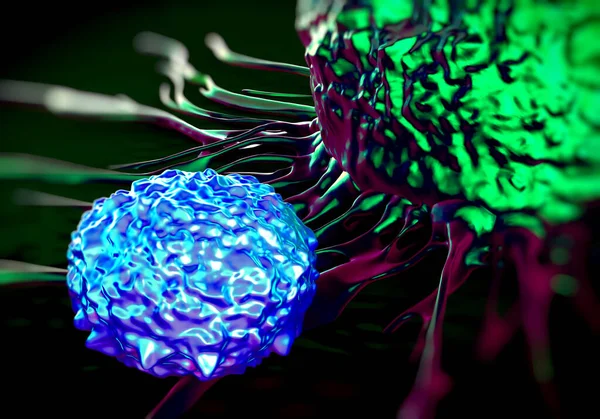
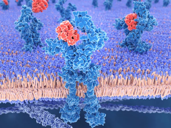
The T-cell Receptor Activates The Immune Response To Antigens In T-lymphocytes. T-cell Receptors (dark Blue), CD4 Molecules (light Blue), Glycolipids (orange). 3d Rendering. Illustration

Image, 3.11MB, 8000 × 6000 jpg
stock image Eosinophil

Eosinophil

Image, 4.42MB, 3872 × 2592 jpg
stock image Lymphoblast

Lymphoblast

Image, 5.36MB, 3872 × 2592 jpg
stock image Ribosome translating mRNA into a polypeptide chain

Ribosome Translating MRNA Into A Polypeptide Chain

Image, 8.4MB, 8000 × 6000 jpg
stock image lymphocyte

Lymphocyte

Image, 3.98MB, 3872 × 2592 jpg
stock image 3d computer illustration of interleukin 13 and its receptor.IL-13 is a cytokine that plays a central regulator role in IgE synthesis, goblet cell hyperplasia, mucus hypersecretion, airway hyperresponsiveness, fibrosis,  It is a mediator of allergic i

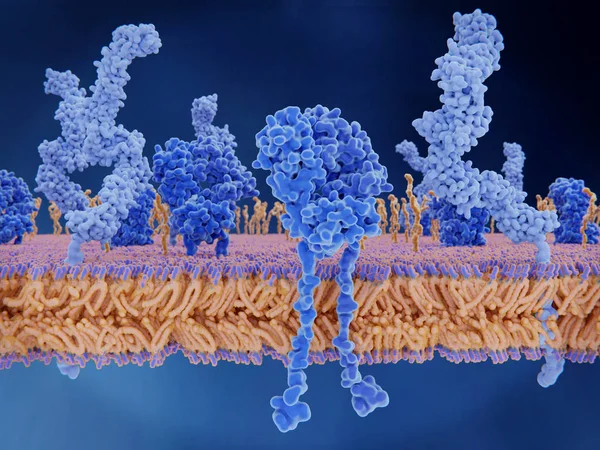
3d Computer Illustration Of Interleukin 13 And Its Receptor.IL-13 Is A Cytokine That Plays A Central Regulator Role In IgE Synthesis, Goblet Cell Hyperplasia, Mucus Hypersecretion, Airway Hyperresponsiveness, Fibrosis, It Is A Mediator Of Allergic I

Image, 5.59MB, 8000 × 6000 jpg
stock image Monocyte

Monocyte

Image, 5.72MB, 3872 × 2592 jpg
stock image Neutrophil

Neutrophil

Image, 5.98MB, 3872 × 2592 jpg
stock image lymphocyte

Lymphocyte

Image, 3.67MB, 3872 × 2592 jpg
stock vector immune system icon set

Immune System Icon Set

Vector, 2.7MB, 4167 × 4167 eps
stock image Neutrophil

Neutrophil

Image, 4.19MB, 3872 × 2592 jpg
stock vector Immune or Lymphatic system. Human's internal lymphoid organs for immune response. Peyer's patch in small intestine, tonsils, Bone marrow, Appendix, Spleen, Thymus, Tonsils, and Lymph nodes. Immunology. Infographics for education. vector illustration.

Immune Or Lymphatic System. Human's Internal Lymphoid Organs For Immune Response. Peyer's Patch In Small Intestine, Tonsils, Bone Marrow, Appendix, Spleen, Thymus, Tonsils, And Lymph Nodes. Immunology. Infographics For Education. Vector Illustration.

Vector, 4.27MB, 4444 × 4444 eps
stock image Neutrophil

Neutrophil

Image, 3.4MB, 2592 × 3872 jpg
stock image red blood cells with white blood cells background.

Red Blood Cells With White Blood Cells Background.

Image, 8.68MB, 5496 × 3636 jpg
stock image Bacteria attacking immune system

Bacteria Attacking Immune System

Image, 1.68MB, 3840 × 2160 jpg
stock image Atypical lymphocyte or Transform lymphocyte.

Atypical Lymphocyte Or Transform Lymphocyte.

Image, 4.02MB, 3872 × 2592 jpg
stock vector Myasthenia gravis. An autoimmune disease of the neuromuscular junction when antibodies block or destroy nicotinic acetylcholine receptors (AChR) at the junction between the nerve and muscle.

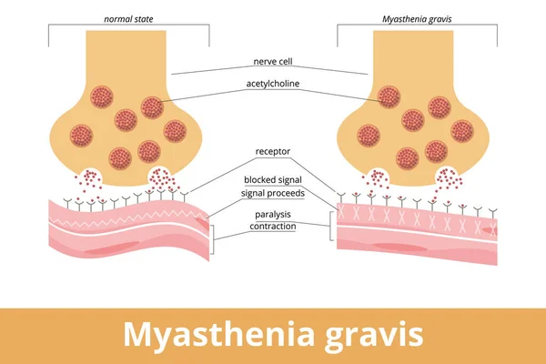
Myasthenia Gravis. An Autoimmune Disease Of The Neuromuscular Junction When Antibodies Block Or Destroy Nicotinic Acetylcholine Receptors (AChR) At The Junction Between The Nerve And Muscle.

Vector, 7.31MB, 6250 × 4167 eps
stock vector Immune response

Immune Response

Vector, 1.12MB, 2000 × 2268 eps
stock image giant platelet

Giant Platelet

Image, 6.35MB, 3872 × 2592 jpg
stock vector Vector illustration of the response of the immune system to pollen allergy

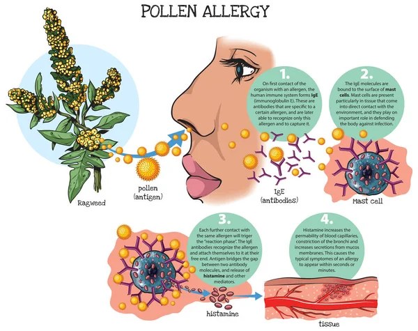
Vector Illustration Of The Response Of The Immune System To Pollen Allergy

Vector, 14.34MB, 3782 × 3000 ai
stock image Bacteria attacking immune system

Bacteria Attacking Immune System

Image, 1.88MB, 3840 × 2160 jpg
stock image The basophils are a type of white blood cell on the black background 3d rendering

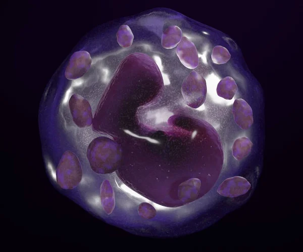
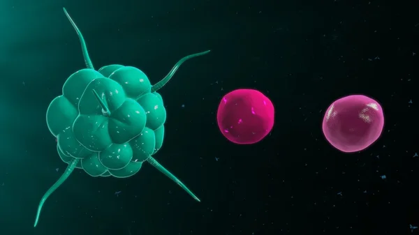
The Basophils Are A Type Of White Blood Cell On The Black Background 3d Rendering

Image, 0.27MB, 2400 × 2000 jpg
stock image lymphocyte

Lymphocyte

Image, 3.98MB, 3872 × 2592 jpg
stock vector Mast cell: synthesis. Due to antigen activation, mast cells produce prostaglandins, leukotrienes, histamine, and cytokines. Visualization of mast cell products during an allergic reaction.

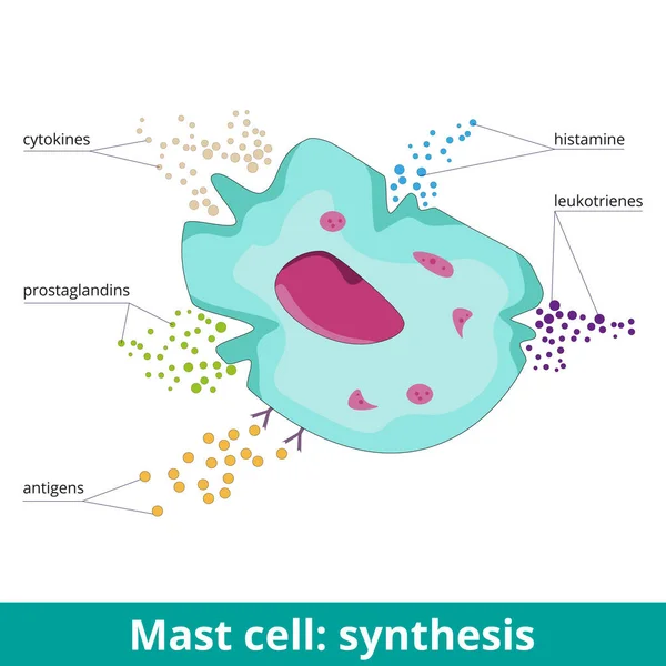
Mast Cell: Synthesis. Due To Antigen Activation, Mast Cells Produce Prostaglandins, Leukotrienes, Histamine, And Cytokines. Visualization Of Mast Cell Products During An Allergic Reaction.

Vector, 5.85MB, 6250 × 6250 eps
stock image metamyelocyte

Metamyelocyte

Image, 4.24MB, 3872 × 2592 jpg
stock image Neutrophil

Neutrophil

Image, 4.28MB, 3872 × 2592 jpg
stock image Segmented neutrophils

Segmented Neutrophils

Image, 5.06MB, 3872 × 2592 jpg
stock image T cells and B cells in immune system

T Cells And B Cells In Immune System

Image, 1.47MB, 3840 × 2160 jpg
stock image Monocyte

Monocyte

Image, 3.91MB, 3872 × 2592 jpg
Previous << Page 2 >> Next