
Stock image Inhibitory

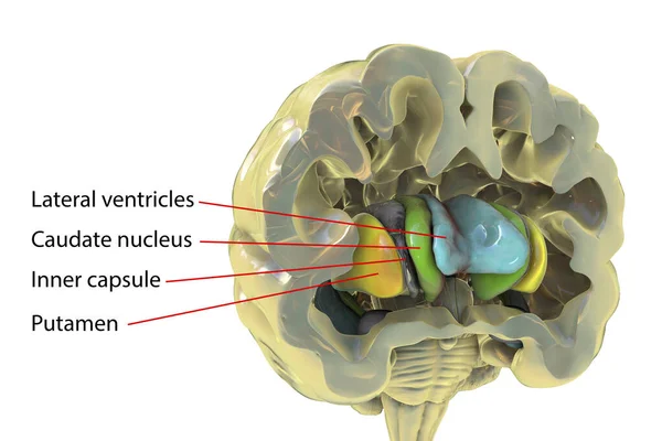
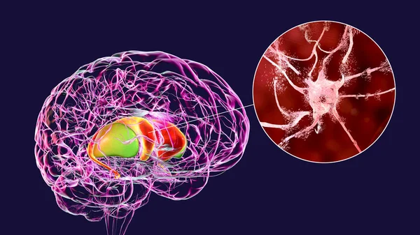
Human Brain Anatomy, Basal Ganglia. 3D Illustration Showing Caudate Nucleus (green), Putamen (yellow), And Lateral Ventricles (blue)
Image, 6.25MB, 6252 × 4168 jpg
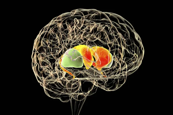
Dorsal Striatum In The Human Brain, 3D Illustration. It Is A Nucleus In The Basal Ganglia, Consists Of The Caudate Nucleus (red) And The Putamen (green), Is A Component Of The Motor And Reward Systems
Image, 6.21MB, 6000 × 4000 jpg

Executive Function Or Cognitive Control, Vector Illustration Outline Diagram
Vector, 6.05MB, 4000 × 4000 eps
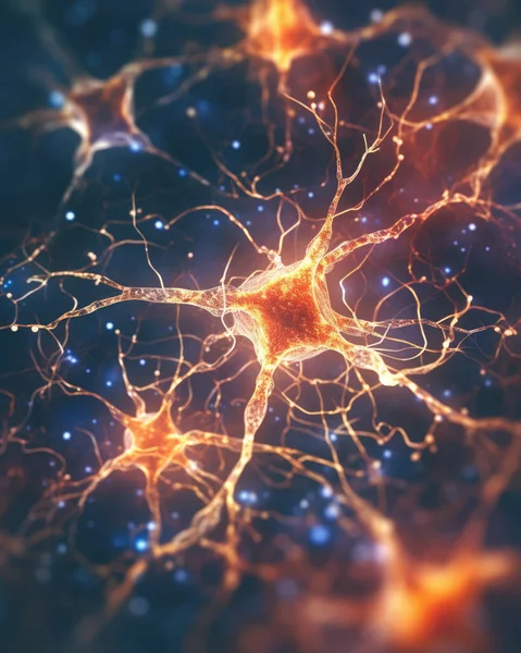
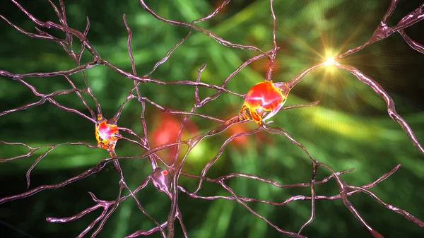
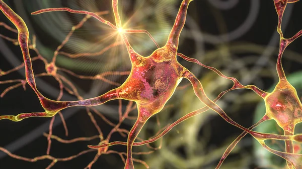
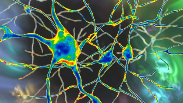
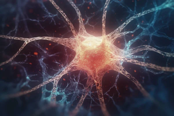
Neuron Conceptual Image Of Human Nervous System. 3D Illustration Of Neurons With Vivid Colors.
Image, 16.7MB, 3880 × 4850 jpg
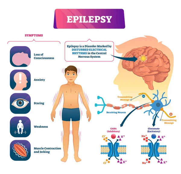
Epilepsy Vector Illustration. Labeled Sick CNS Disorder Educational Scheme.
Vector, 6.34MB, 4200 × 4035 eps
Dorsal Striatum Highlighted In Human Brain And Close-up View Of Degrading Neurons Of Dorsal Striatum Seen In Huntington's Disease, 3D Illustration
Image, 16.37MB, 8672 × 4878 jpg

Adaptive Immune Response, Highlighting The Interaction Between NK Cells And Healthy Versus Infected Cells Diagram Hand Drawn Schematic Raster Illustration. Medical Science Educational Illustration
Image, 3.4MB, 6000 × 4500 jpg
Neurons Of Dorsal Striatum, 3D Illustration. The Dorsal Striatum Is A Nucleus In The Basal Ganglia, Degrading Of Its Neurons Plays A Crucial Role In The Development Of Huntington's Disease
Image, 10.79MB, 7200 × 4050 jpg

Attention Deficit Hyperactivity Disorder Written On A Book And Diagnosis Form.
Image, 8.02MB, 6000 × 4000 jpg
Neurons Of Dorsal Striatum, 3D Illustration. The Dorsal Striatum Is A Nucleus In The Basal Ganglia, Degrading Of Its Neurons Plays A Crucial Role In The Development Of Huntington's Disease
Image, 12.53MB, 7200 × 4050 jpg

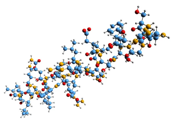
Structure Of Human Interleukin-11, 3D Cartoon Model Isolated, White Background
Image, 2.35MB, 6000 × 4000 jpg
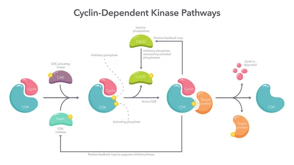
Cyclin Dependent Kinase Activation Pathway Science Vector Illustration Infographic
Vector, 0.45MB, 8333 × 4688 ai

No Skiing Outside Ski Track Prohibition Sign In Krasnaya Polyana Resort Against Snowy Aibga Mountain Peak Background. Sochi. Russia, Caucasus
Image, 5.26MB, 4000 × 2667 jpg
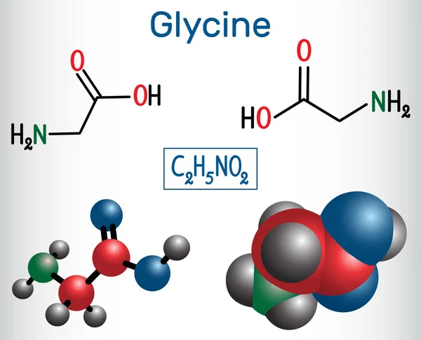
Glycine, Gly Or G , Is The Amino Acid. Structural Chemical Formula And Molecule Model
Vector, 5.22MB, 5000 × 4048 eps
Vector Round Banner Cognitive Skills With Icons. Editable Stroke. Attention Recognition, Mental Health, Working Memory, Psychology Pattern. Cognitive Control Flexibility And Speed Of Thinking, Autism.
Vector, 0.89MB, 4000 × 4000 eps

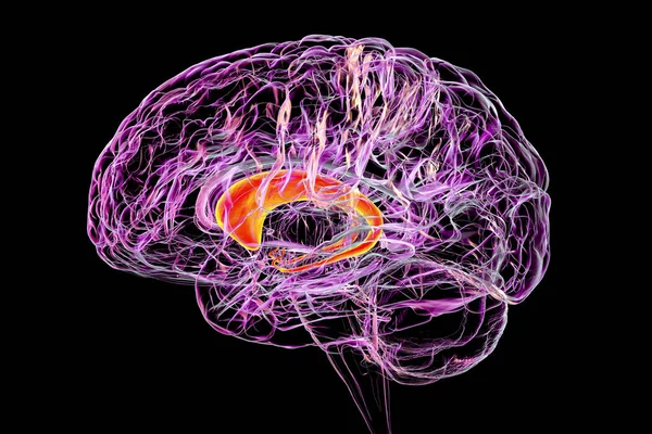
Coronal Section Of A Healthy Brain Showing Normal Anatomy Of Basal Baglia And Ventricles, 3D Illustration
Image, 4.88MB, 6000 × 4000 jpg

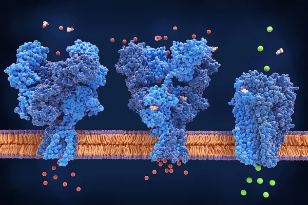
Activation Of The GABA B Receptor By Baclofen. GABA B Receptors Are G Protein-coupled Receptors. Binding Of An Agonist (baclofen, Red) Leads To A G-protein Coupled C-AMP Signaling Pathway. Source: PDB Entries 7eb2, 6r3q,.
Image, 9.97MB, 8000 × 6000 jpg

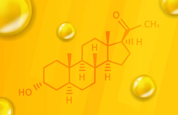
Allopregnanolone Chemical Formula. Allopregnanolone 3D Realistic Chemical Molecular Structure
Vector, 2.75MB, 6200 × 4000 eps
Neurons Of Dorsal Striatum, 3D Illustration. The Dorsal Striatum Is A Nucleus In The Basal Ganglia, Degrading Of Its Neurons Plays A Crucial Role In The Development Of Huntington's Disease
Image, 8.02MB, 7200 × 4050 jpg
Neurons Of Dorsal Striatum, 3D Illustration. Dorsal Striatum Is A Nucleus In The Basal Ganglia, Degrading Of Its Neurons Plays Crucial Role In Development Of Huntington's Disease
Image, 12.86MB, 7200 × 4050 jpg

Adaptive Immune Response, Highlighting The Interaction Between NK Cells And Healthy Versus Infected Cells Diagram Hand Drawn Schematic Vector Illustration. Medical Science Educational Illustration
Vector, 0.62MB, 5000 × 3750 eps

Neurons Of Dorsal Striatum, 3D Illustration. Dorsal Striatum Is A Nucleus In The Basal Ganglia, Degrading Of Its Neurons Plays Crucial Role In Development Of Huntington's Disease
Image, 8.32MB, 7200 × 4050 jpg

Brain Dorsal Striatum Anatomy, 3D Illustration. The Dorsal Striatum Consists Of The Caudate Nucleus (orange) And The Putamen (blue). Amygdala Is Colored In Red. Front View
Image, 5.28MB, 6000 × 4000 jpg

Caudate Nuclei Highlighted In Human Brain, 3D Illustration. The Caudate Nucleus Is A Component Of The Basal Ganglia, It Is Associated With Motor Processes And Plays Role In Parkinson's Disease
Image, 9.68MB, 6000 × 4000 jpg
3D Image Of Secretin Skeletal Formula - Molecular Chemical Structure Of Water Homeostasis Hormone Isolated On White Background
Image, 4.9MB, 7000 × 4922 jpg
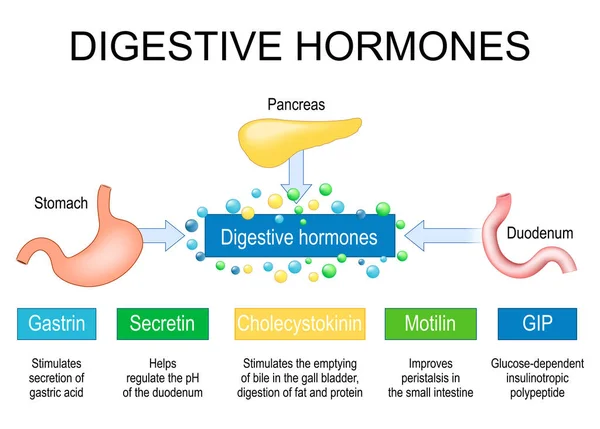
Digestive Hormones. Human Internal Organs Pancreas, Stomach And Duodenum Like Part Of Endocrine System. Gastrointestinal Hormones Gastrin, Cholecystokinin, Secretin, Gastric Inhibitory Peptide And Motilin. Vector Illustration
Vector, 4.94MB, 5000 × 3580 eps


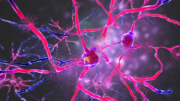
Neuron Conceptual Image Of Human Nervous System. 3D Illustration Of Neurons With Vivid Colors.
Image, 10.88MB, 4800 × 3200 jpg
Page 1 >> Next