Home
Images
Vectors
Videos
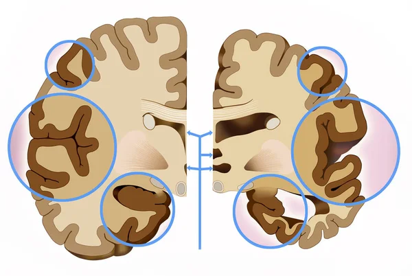
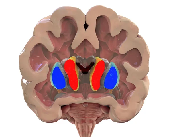
Stock image Coronal section of a healthy brain showing normal anatomy of basal baglia and ventricles, 3D illustration
Published:
May.24, 2021 13:03:01
Author:
katerynakon
Views:
22
Downloads:
1
File type:
image
/
jpg
File size:
4.88 MB
Orginal size:
6000
x
4000
px
Available sizes:
S
(500 x 333)
M
(1000 x 667)
L
(2000 x 1333)
XL
(6000 x 4000)
EL0
(6000 x 4000)
Level:
silver
Download Image
Image topics:
Illustration
Isolated
Human
Healthy
Head
Medicine
Medical
3d
Body
Learning
Intelligence
Science
Organ
Control
Cortex
Mind
Motor
Nucleus
Anatomy
Normal
Central
Nervous
Disorder
Neurology
Anatomical
Dorsal
Associative
Nuclei
Putamen
Neuro
Basal
Claustrum
Inhibitory
White Background
Caudate
Ganglia
Striatum
Grey Matter
Coronal Section
Lateral Ventricle
Corpus Callosum
Parkinson Disease
White Matter
Corpus Striatum
Caudate Nucleus
Globus Pallidum
Similar stock images
Coronal Section Of A Brain Of A Person With Huntington's Disease Showing Enlarged Anterior Horns Of The Lateral Ventricles, Degeneration And Atrophy Of The Dorsal Striatum, 3D Illustration
6000 × 4000
Coronal Sections Of A Healthy Brain And A Brain In Huntington's Disease Showing Enlarged Anterior Horns Of The Lateral Ventricles, Degeneration And Atrophy Of The Dorsal Striatum, 3D Illustration
10000 × 5000
This Illustration Shows The Comparison Of Two Halves Of The Brain, A Healthy Half And Another With Alzheimer's, In This We See That The Cerebral Cortex Contracts, This Brain Reduction Causes Damage To The Brain Used To Think, Plan And Remem
3921 × 2629
Mid Sagittal Section Of The Human Brain Showing The Hypothalamus. .
3828 × 3020
Realistic Human Sick Brain Isolated On White Background.
5000 × 5000
Coronal Section Of A Healthy Brain Showing Normal Anatomy Of Basal Baglia And Ventricles, 3D Illustration. Non-labelled Version Of The Image
6250 × 5000
Human Brain
3460 × 3930