
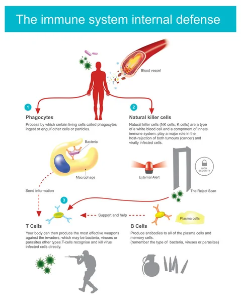
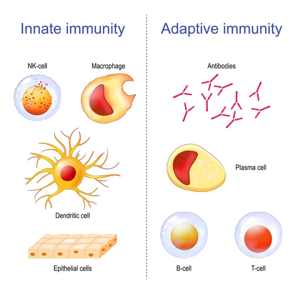
Stock image Innate Immunity

Innate And Adaptive Immune System. Immunology Infographic With Cell Types. Immunity Response, Antibody Activation, Lymphocytes Vector Scheme
Vector, 2.54MB, 5000 × 5000 eps
Immune System Cells Vector Illustration. Labeled Educational Division Scheme.
Vector, 14.1MB, 3938 × 4500 eps
Makrophage, Type Of White Blood Cell, Of The Immune System, That Engulfs And Digests Cellular Debris, Foreign Substances, Microbes, Cancer Cells
Image, 9.6MB, 4000 × 3000 jpg

Macrophages Approaching Bacteria (bacilli), 3D Rendering. Illustration
Image, 2.49MB, 8000 × 6000 jpg
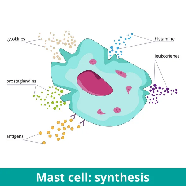
Mast Cell: Synthesis. Due To Antigen Activation, Mast Cells Produce Prostaglandins, Leukotrienes, Histamine, And Cytokines. Visualization Of Mast Cell Products During An Allergic Reaction.
Vector, 5.85MB, 6250 × 6250 eps
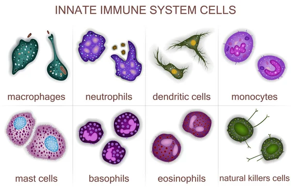
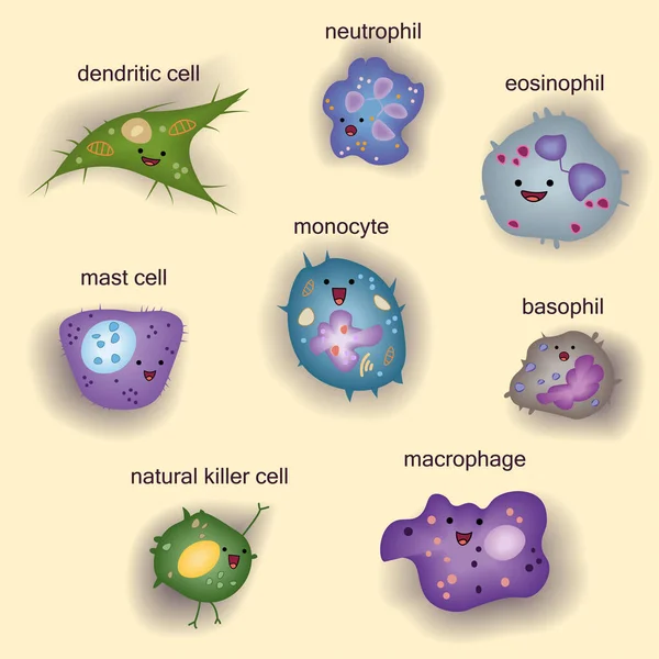
Set Of Innate Immune System Cells, Cartoon Cute Funny Vector Illustration
Vector, 7.73MB, 4792 × 4792 eps
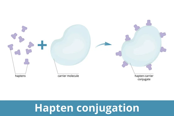
Hapten Conjugation. Haptens Are Small Molecules That Elicit An Immune Response When Attached To A Large Carrier (protein). Formation Of Immunogenic Carrier Proteins.
Vector, 5.74MB, 6250 × 4167 eps
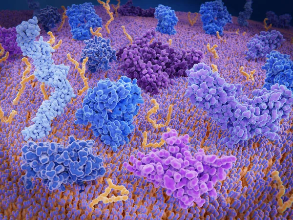
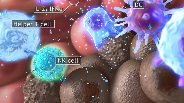
Dendritic Cells Present Antigens (green) To Lymphocytes Through Their Membran Bound MHC-molecules (violet). CD4 Molecules (light Blue) Bind To Other Portions Of The MHC, Strengthening The Interaction.
Image, 10.24MB, 8000 × 6000 jpg
3d Computer Illustration Of A Dendritic Cell. They Areantigen-presenting Cells Of The Immune System. Their Main Function Is To Process Antigen Material And Present It On The Cell Surface To The T Cells Of The Immune System. They Are Messengers Betwe
Image, 5.2MB, 8000 × 6000 jpg

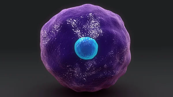
Leukocyte, White Blood Cell Seen Under A Microscope. Cell. 3d Rendering
Image, 3.46MB, 4724 × 4724 jpg
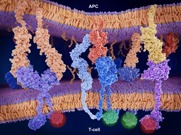
Interaction Of MHC-II (red) With The T-cell Receptor (blue) And CD4 (light Blue) And B7-1 (orange) With CD-28 (dark Blue) Activates T-cells While The Interaction Of P7-1 With CTLA-4 (violett) And PD-L1 (yellow) With PD-1 Deactivates T-cells
Image, 10.65MB, 8000 × 6000 jpg
PD-1 (red) Extends From The Surface Of A T-cell And Interacts With The Ligand Protein PD-L1 (yellow) From A Antigen Presenting Cell. Although The T-cell Has Been Activated Through The Interaction Of A T-cell Receptor (blue) And A MHC Protein (viole
Image, 18.32MB, 8000 × 6000 jpg

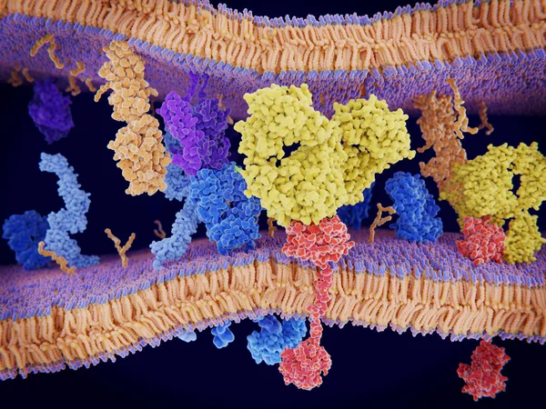
Interactions Of MHC-II With The T-cell Receptor And CD4 And B7-1 With CD-28 Activates T-cells While The Interactions Of P7-1 With CTLA-4 And PD-L1 With PD-1 Deactivates T-cells.
Image, 10.7MB, 8000 × 6000 jpg
Cancer Cells Express PD-L1 (orange) Proteins On Their Surface To Trick The Immune System. The Interaction Of PD-L1 With PD-1 Of T-cells Leads To A Down-regulation Of T-cells. The Antibody (yellow) Blocks This Interaction.
Image, 18.3MB, 8000 × 6000 jpg
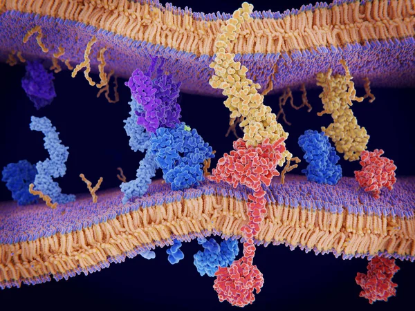
Immunologically Active Proteins On A T-cell. TCR (blue), CD-4 (light Blue), CD-28 (dark Blue), PD-1 (magenta), CTLA-4 (violet), Ca-channel (dark Violet). The T-cell Receptor, CD-4 And CD-28 Activate T-cells, While PD-1 And CTLA-4 Inhibit The Activat
Image, 10.2MB, 8000 × 6000 jpg
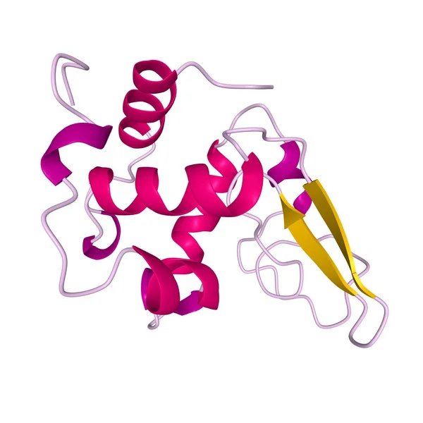
Native Human Lysozyme, 3D Cartoon Model Of The Tertiary Structure With The Elements Of The Secondary Structure Colored, White Background
Image, 1.1MB, 4096 × 4096 jpg
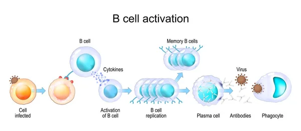
Activation Of B Cell Leukocytes. Transparent Realistic Cells Of Adaptive And Innate Immune System. Vector Poster
Vector, 8.78MB, 6879 × 3000 eps
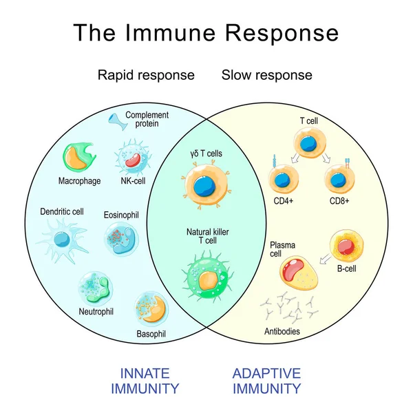
Immune Response. Rapid And Slow Response Of Adaptive And Innate Immunity And Antibody Activation. Cells Of The Immune System. Immunology Infographic. Vector Illustration
Vector, 2.28MB, 4444 × 4444 eps
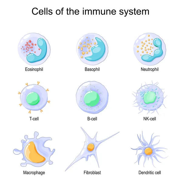
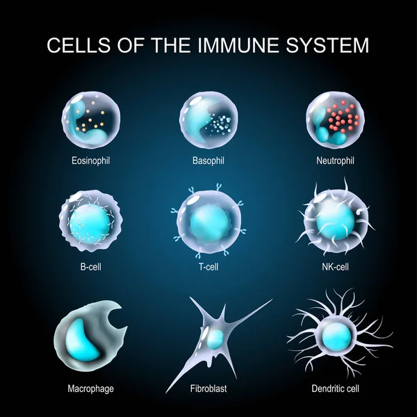
Cells Of The Immune System. White Blood Cells Or Leukocytes Eosinophil, Neutrophil, Basophil, Macrophage, Fibroblast, And Dendritic Cell. Vector Illustration
Vector, 1.73MB, 4444 × 4445 eps
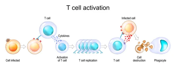
Activation Of Leukocytes. T-cell Encounters Its Cognate Antigen On The Surface Of An Infected Cell. T-cells Direct And Regulate Immune Responses And Attack Infected Or Cancerous Cells. Cell-mediated Immunity. The Adaptive And Innate Immune System. Ve
Vector, 7.72MB, 7736 × 3000 eps
Cells Of The Immune System. White Blood Cells Or Leukocytes Eosinophil, Neutrophil, Basophil, Macrophage, Fibroblast, And Dendritic Cell. Set Of Transparent Realistic Cells On A Dark Background. Vector Illustration
Vector, 5.41MB, 4444 × 4444 eps
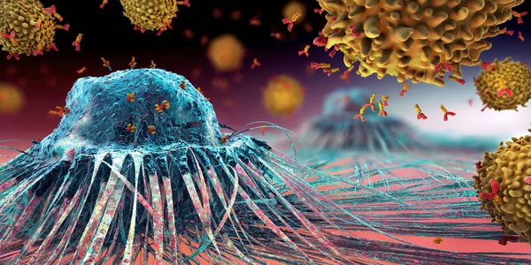
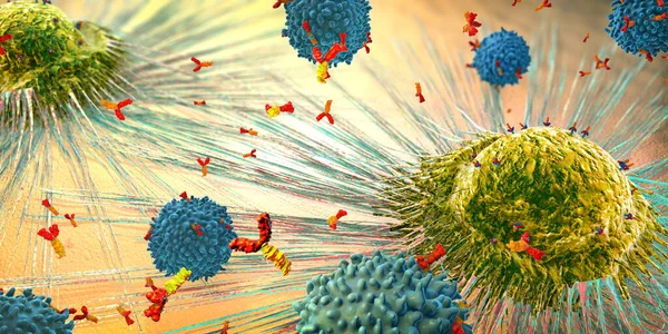
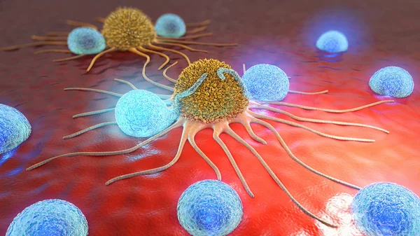
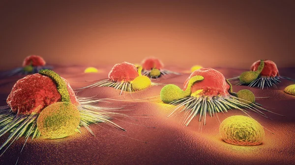
Lymphocytes Cell In The Immune System Reacting And Attacking A Spreading Cancer Cell - 3d Illustration
Image, 22.42MB, 7000 × 3500 jpg
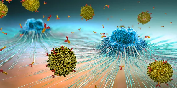
Lymphocytes Cell In The Immune System Reacting And Attacking A Spreading Cancer Cell - 3d Illustration
Image, 18.3MB, 7000 × 3500 jpg
3D Medical Illustration Cytotoxic T Cell And Helper T Cell Destroys The Stress-related Molecule
Image, 1.77MB, 3840 × 2160 jpg

Innate Immunity From Fever And Complement System (protein For Holes In The Plasma Membrane), To Macrophage, NK And Dendritic Cells. Adaptive Immunity From Antibodies And Plasma Cell To B-cell, T Helper, T-killer. Comparison And Difference
Vector, 2.86MB, 4444 × 4444 eps

Lymphocytes Cell In The Immune System Reacting And Attacking A Spreading Cancer Cell - 3d Illustration
Image, 19.24MB, 7000 × 3500 jpg
Medical Science Background, Neutrophils, A Large Group Of Leukocyte Granulocytes, Are Part Of Innate Immunity, The Main Function Is Phagocytosis Of Pathogenic Microorganisms, 3d Rendering
Image, 3.87MB, 2880 × 2880 jpg
Adaptive Immunity: T-cell, Antibodies, Plasma Cell And B-cell. Innate Immunity: Macrophage, Dendritic, Epithelial, And NK Cells. Immunology Infographic. Vector Illustration
Vector, 14.03MB, 4444 × 4444 eps

Innate Immune System. Anatomical Barriers. Man Silhouette With Internal Organs. Blood Brain Barrier Protects The Nervous System From Pathogens. Shedding O
Vector, 12.03MB, 4444 × 4444 eps
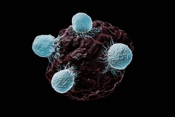
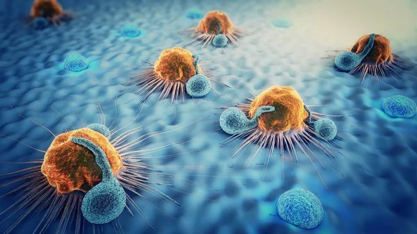
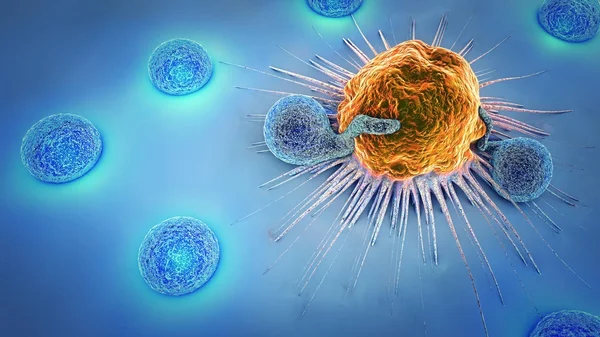
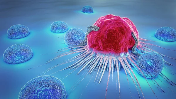
White Blood Cells, T Lymphocytes Or Natural Killers T Attack A Cancerous Or Infected Cell 3D Rendering Illustration Isolated On Black Background. Science, Medicine, Biomedical Research, Immune System, Oncology, Biology Concepts.
Image, 2.24MB, 3500 × 2333 jpg

White Blood Cells Or T Lymphocytes Or Natural Killer T Attack A Cancer Or Tumor Or Infected Cell 3D Rendering Illustration. Oncology, Immune System, Biomedical, Medicine, Science, Biology Concepts.
Image, 3.87MB, 3500 × 2333 jpg


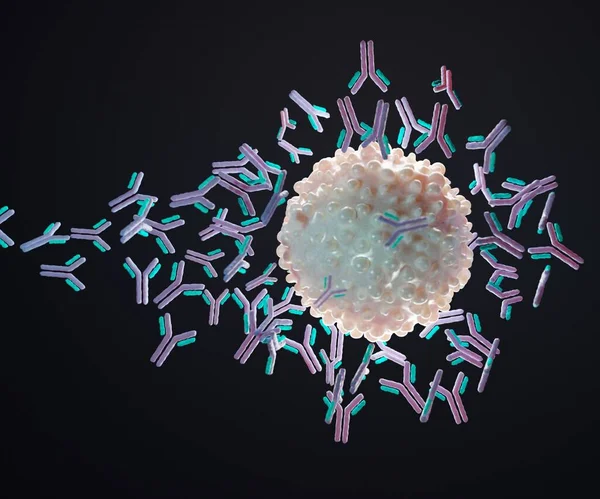
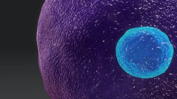
An Antigen Is A Molecule That Initiates The Production Of An Antibody And Causes An Immune Response 3d Rendering
Image, 0.3MB, 2400 × 2000 jpg

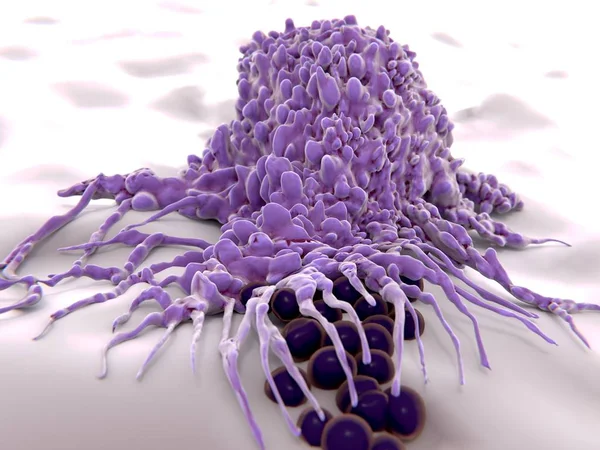
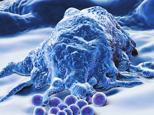
Macrophag Engulfing Bacteria (cocci), 3D Rendering. Macrophages Engulf And Digest Cellular Debris And Pathogens. - Illustration
Image, 2.72MB, 8000 × 6000 jpg

Page 1 >> Next