Home
Images
Vectors
Videos
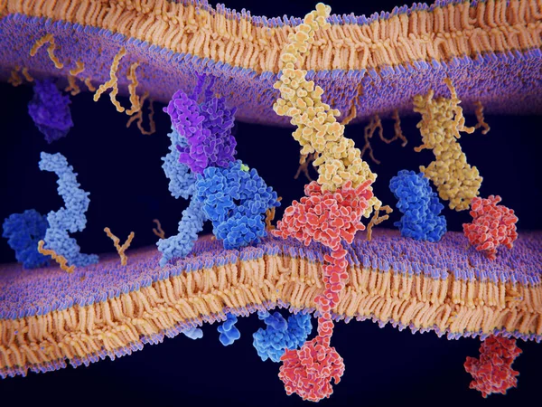
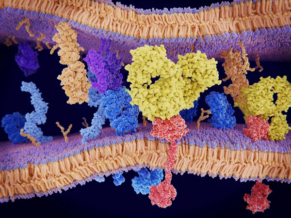
Stock image Cancer cells express PD-L1 (orange) proteins on their surface to trick the immune system. The interaction of PD-L1 with PD-1 of T-cells leads to a down-regulation of T-cells. The antibody (yellow) blocks this interaction.
Published:
Mar.15, 2019 08:15:28
Author:
animaxx3d
Views:
70
Downloads:
7
File type:
image
/
jpg
File size:
18.3 MB
Orginal size:
8000
x
6000
px
Available sizes:
S
(500 x 375)
M
(1000 x 750)
L
(2000 x 1500)
XL
(8000 x 6000)
EL0
(8000 x 6000)
Level:
bronze
Download Image
Image topics:
Natural
Protein
Medicine
Illness
Disease
Cancer
Immunization
Immunity
Molecules
Receptor
Antigen
Lymphocyte
Metastasis
Peptide
Human Cell
White Blood Cell
Cytotoxic
Immune System
Cancer Prevention
Cell Membrane
Interferon
Major Histocompatibility Complex
White Cell
Cancer Therapy
Immune Defense
Signal Transduction
Killer Cell
Mhc Complex
Co Stimulation
Dendritic Cell
Adaptive Immunity
Innate Immunity
Antigen Presenting Cells
Antigen Presentation
Mhc Class
Pd 1
Pd L1
Accessory Cell
Down Regulation
T Cell Receptor
Helper T Cell
Programmed Cell Death Protein
Signal Cascade
Malignant Cell
Cytotoxic T Cell
Similar stock images
PD-1 (red) Extends From The Surface Of A T-cell And Interacts With The Ligand Protein PD-L1 (yellow) From A Antigen Presenting Cell. Although The T-cell Has Been Activated Through The Interaction Of A T-cell Receptor (blue) And A MHC Protein (viole
8000 × 6000
Activation Of The Immune Response To An Antigene (green) Through The Complex Between A T-cell Receptor (dark Blue), An MHC II-antigen (violet) And A CD4 Protein (light Blue). 3d Rendering. Illustration
8000 × 6000
The T-cell Receptor Activates The Immune Response To Antigens In T-lymphocytes. T-cell Receptors (dark Blue), CD4 Molecules (light Blue), Glycolipids (orange). 3d Rendering. Illustration
8000 × 6000