
Stock image Intraventricular
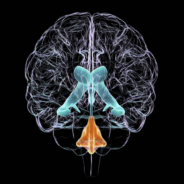
A 3D Scientific Illustration Depicting Isolated Enlargement Of The Fourth Brain Ventricle, Front View.
Image, 12.67MB, 6000 × 6000 jpg
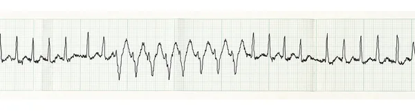
ECG With Paroxysm Of Atrial Fibrillation And Transient Disturbances Of Intraventricular Conduction
Image, 1.92MB, 3900 × 1050 jpg

In Complete Left Bundle Branch Block, The Conduction Of The LBB Can Be Completely Interrupted Or Can Still Be Conducted, But It Is Delayed By At Least 45ms Than The RBB.
Image, 10.8MB, 10000 × 5497 jpg

Intracranial Pressure. ICP Monitoring. Cross Section Of A Human Brain. Localizations Of Pressure Probes Or Catheters. Epidural, Subdural, Parenchymal, Intraventricular. Surgical Techniques In Trauma. Vector Illustration
Vector, 9.04MB, 4444 × 4444 eps
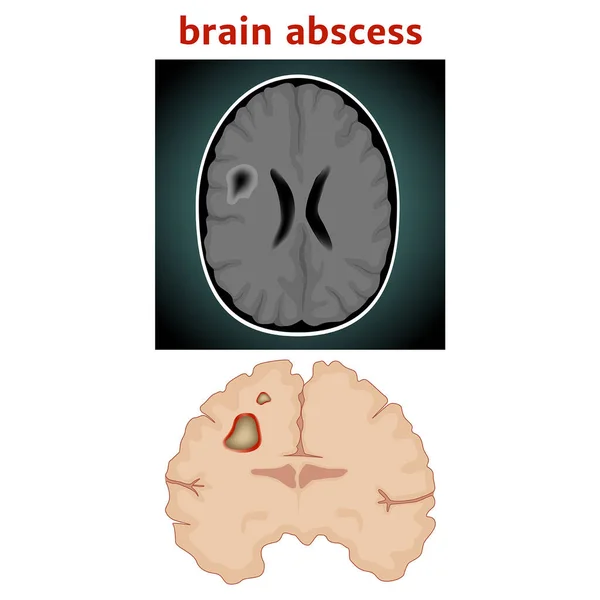
Brain Abscess. Limited Purulent Fusion Of The Substance. Medical Poster. Vector Illustration
Vector, 8.68MB, 2000 × 2000 eps

A 2:1 Left Bundle Branch Block Is Considered When Complete Left Bundle Branch Block Alternates With Normal QRS Complexes And The PR Interval Is Fixed.
Image, 5.72MB, 10000 × 3162 jpg

During Left Posterior Fascicular Block, The ECG Showed Right Axis Deviation. The QRS Wave In Leads I And AVL Was RS Wave, And The Duration Of QRS Wave Was Less Than 120 Ms.
Image, 30.53MB, 10000 × 11472 jpg

ECG Tape With Paroxysm Of Atrial Fibrillation And Restoration Of Sinus Rhythm
Image, 12.28MB, 7000 × 1178 jpg
Page 1 >> Next