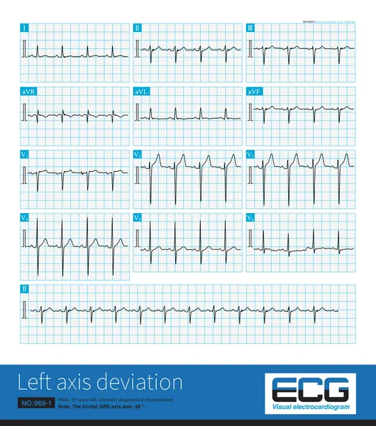
Stock image During left posterior fascicular block, the ECG showed right axis deviation. The QRS wave in leads I and aVL was rS wave, and the duration of QRS wave was less than 120 ms.

Published: Aug.04, 2022 16:40:58
Author: asia11m
Views: 15
Downloads: 1
File type: image / jpg
File size: 30.53 MB
Orginal size: 10000 x 11472 px
Available sizes:
Level: beginner