
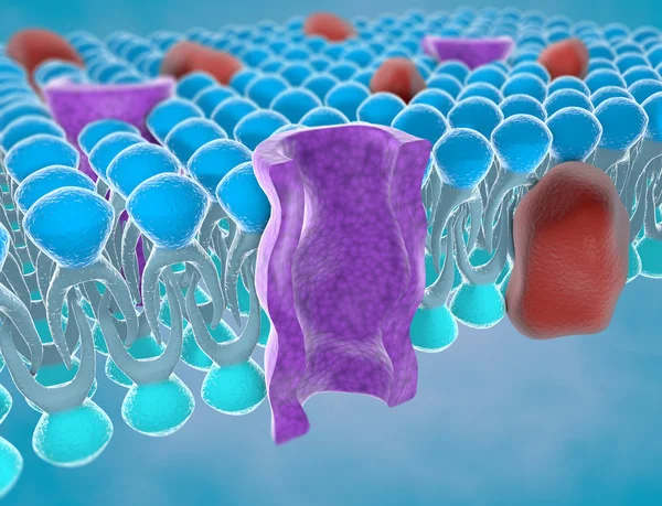
Stock image Ion Channel
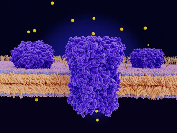
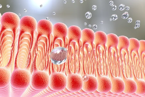
Chloride Channels Conduct Chloride Anions Across Cell Membranes, Regulating The Electrical Excitation Of Skeletal Muscles And The Flow Of Salt And Water Across Epithelial Barriers. They Also Play An Importante Role In Regulation Of PH, Volume Homeos
Image, 5.62MB, 8000 × 6000 jpg
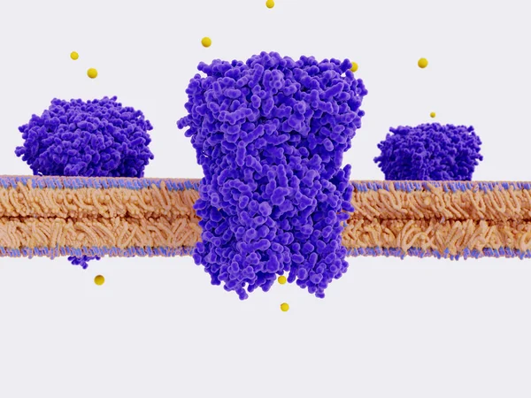
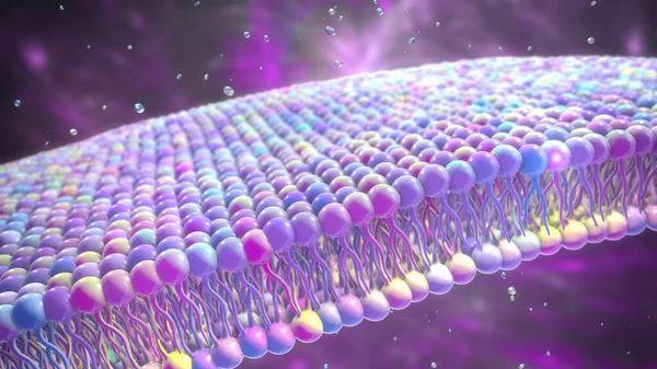
Chloride Channels Conduct Chloride Anions Across Cell Membranes, Regulating The Electrical Excitation Of Skeletal Muscles And The Flow Of Salt And Water Across Epithelial Barriers. They Also Play An Importante Role In Regulation Of PH, Volume Homeos
Image, 4.56MB, 8000 × 6000 jpg
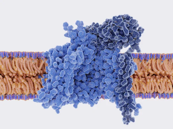
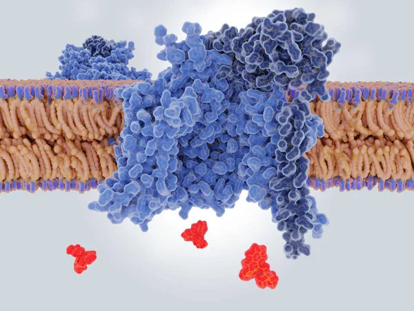
Antidepressive Drug (amitriptilyne) Binding To And Blocking A Sodium Channel
Image, 8.37MB, 8000 × 6000 jpg
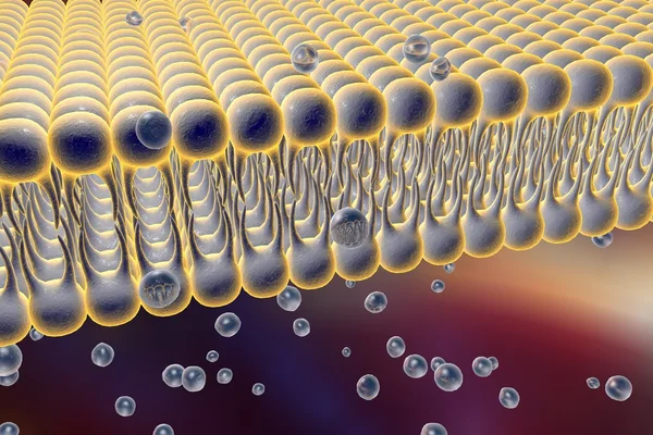
Chloride Channels Conduct Chloride Anions Across Cell Membranes, Regulating Electrical The Excitation Skeletal Muscle And The Flow Of Salt And Water Across Epithelial Barriers. They Also Play An Importante Role In Regulation Of PH, Volume Homeostasi
Image, 29.73MB, 8000 × 6000 jpg
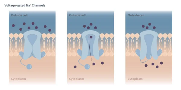
Neuronal Charged Membranes. Voltage-gated Ion Channels Are Closed At The Resting Potential And Open In Response To Changes In Membrane Voltage.
Vector, 7.67MB, 8334 × 4167 eps
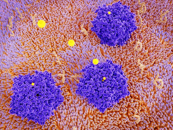
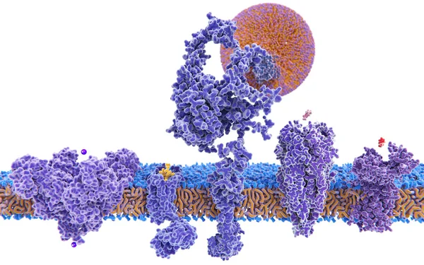
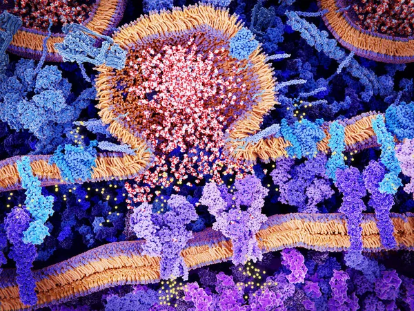
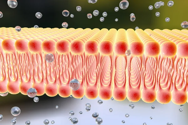
Membrane Proteins (violett), Glycolipids (yellow) And Several Ligands Of The Proteins
Image, 7.89MB, 8000 × 6000 jpg

The Opioid Receptors Display A Role In Modulating Pain Perception; Opioid Agonists Are Therefore Potent Analgesics. They Appear Mainly In The Brain And Spinal Cord. The Endogenous Opioids Are Enkephalin, Dynorphin, Endorphin And Nociceptin.
Image, 7.96MB, 8000 × 6000 jpg

During The Onset Of Variant Angina Pectoris, ECG Is Divided Into Non Fusion Wave, Partial Fusion Wave And Complete Fusion Wave According To The Fusion Degree Of QRS Wave, ST Segment And T Wave.
Image, 7.94MB, 10000 × 6537 jpg
5 Membrane Proteins With Their Ligands: (left To Right) Potassium Channel, Delta-opioid Receptor, LDL Receptor, Acetylcholine Receptor, Histamine Receptor,
Image, 3.8MB, 8000 × 5000 jpg
5 Membrane Proteins With Their Ligands: (left To Right) Potassium Channel, Delta-opioid Receptor, LDL Receptor, Acetylcholine Receptor, Histamine Receptor.
Image, 4.48MB, 8000 × 5000 jpg

Taste Bud With Receptor Cells. Types Of Taste Receptors. Cell Membrane And Ion Channels For Sour, Salty, Sweet, Umami. This Diagram Above Depicts The Signal Transduction Pathway Of The Different Taste.
Vector, 2.83MB, 6105 × 6105 eps
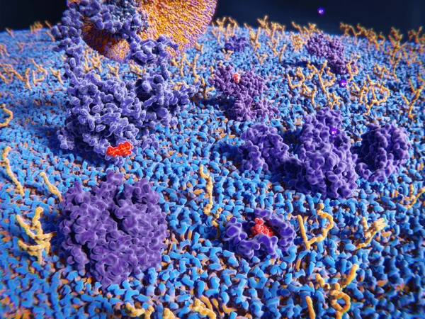
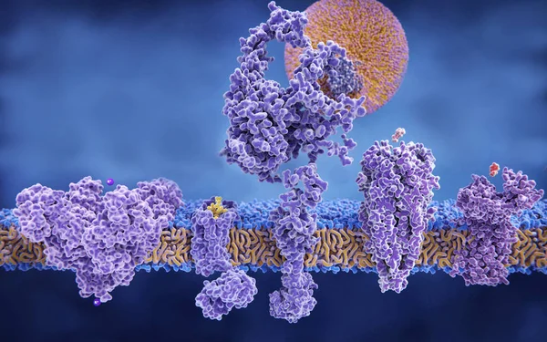
A Cell Membrane In Fish Eye Perspective. Receptors: Opioid Receptor, LDL Receptor And Acetyl Choline Receptor. Channel Proteins: Chloride Channel And Acetat Permease. The Glycolipids Are Depicted In Bluish Green.
Image, 15.26MB, 8000 × 6000 jpg

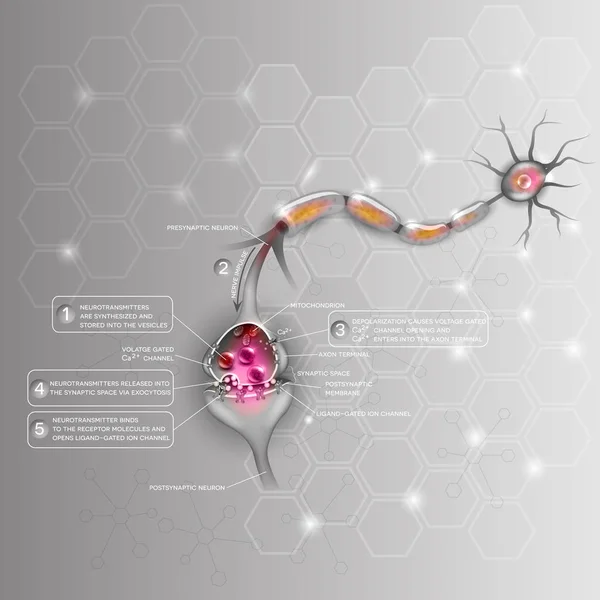
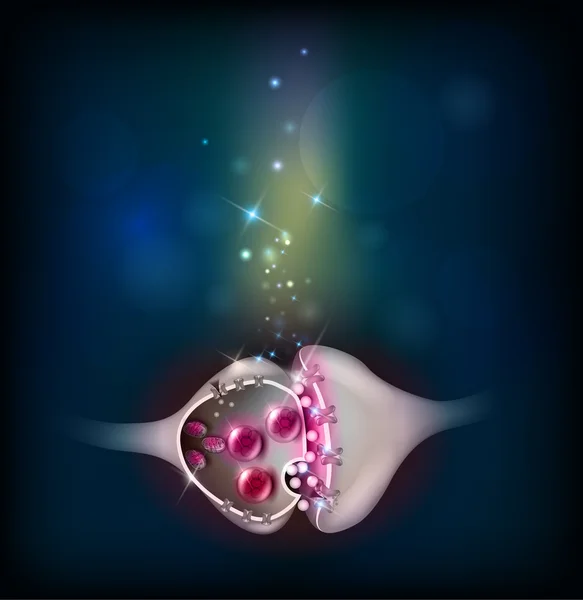
Synapse. Visualization Of Synapse Structure While Passing An Electrical Or Chemical Signal (nervous Impulses) To Another Neuron, Including Mitochondria, Synapse Vesicle, And Ion Channels.
Vector, 6.7MB, 6250 × 6250 eps


Ligand-dependent Ion Channel: Attachment Of A Particular Molecule Causes The Channel To Open.
Image, 1.14MB, 3020 × 4229 jpg

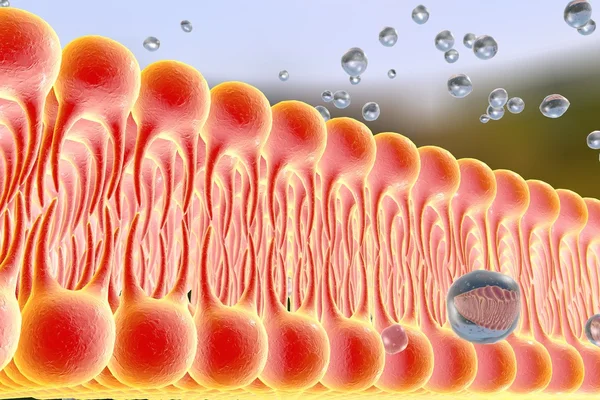
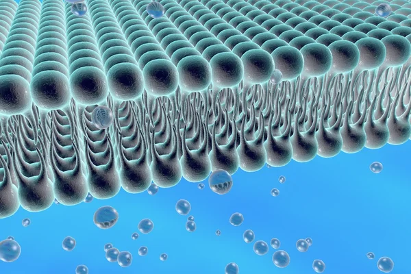
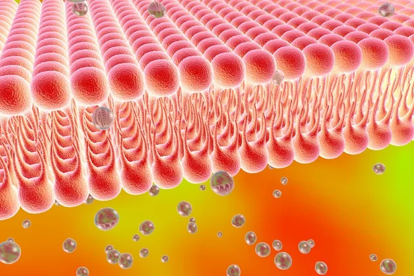
Depolarization: Phospholipid Membrane With NA + And K + Ion Channels.
Image, 2.27MB, 6600 × 5452 jpg

Passive Vs Active Cell Transport. Vector Illustration. Didatic Illustration.
Vector, 0.66MB, 5500 × 4500 ai
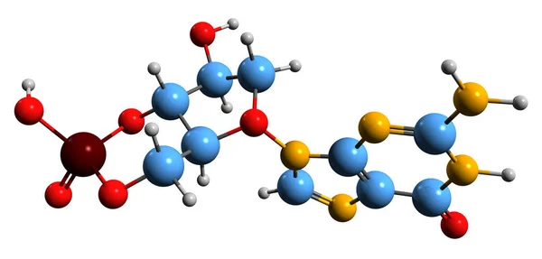
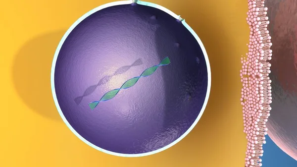
3D Image Of Cyclic Guanosine Monophosphate Skeletal Formula - Molecular Chemical Structure Of Second Messenger CGMP Isolated On White Background
Image, 3.96MB, 8514 × 4080 jpg
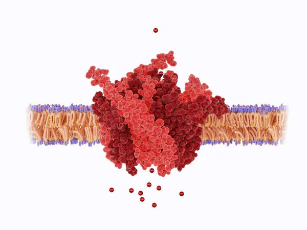
The Calcium Channel Is Composed Of A Hexameric Assembly Or Orai Subunits Around A Central Ion Pore. The Channel Shows Selective Permeability To Calcium Ions.
Image, 3.65MB, 8000 × 6000 jpg


Depolarization: Phospholipid Membrane With NA + And K + Ion Channels.
Image, 2.35MB, 6600 × 5452 jpg


Vector Illustration Of An Example Of Active Transport In Animal Cells - Sodium Potassium Pump.
Vector, 24.69MB, 3000 × 2254 eps


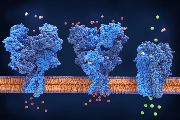
The Neurotransmitter Glutamate Is Transported By Synaptic Vesibles To The Presynaptic Membrane. Calcium Channels Trigger The Neurotransmitter Reslease Into The Inter Synaptic Cleft. Glutamate Binds To The NMDA (left) And AMPA Receptors.
Image, 38.79MB, 8000 × 6000 jpg
Bangkok, Thailand - 26 Mar 2022, The Battery Model For Phantom 4 Pro In Thailand On White Background, It Is Ready For Resale On The Internet Channel.
Image, 6.52MB, 5916 × 3944 jpg

Page 1 >> Next