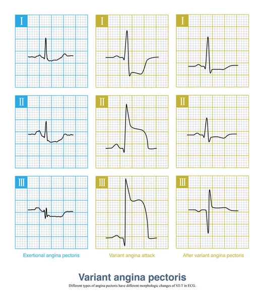
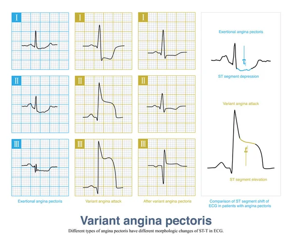
Stock image During the onset of variant angina pectoris, ECG is divided into non fusion wave, partial fusion wave and complete fusion wave according to the fusion degree of QRS wave, ST segment and T wave.

Published: Jul.18, 2022 07:28:18
Author: asia11m
Views: 24
Downloads: 1
File type: image / jpg
File size: 7.94 MB
Orginal size: 10000 x 6537 px
Available sizes:
Level: beginner