
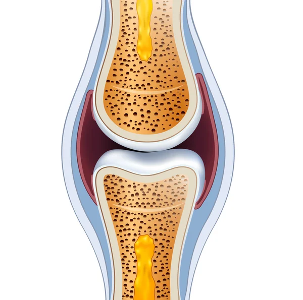
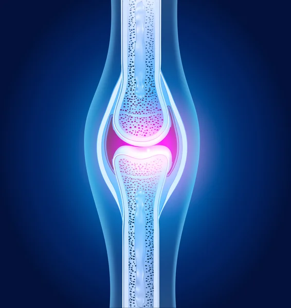
Stock image Joint Cavity

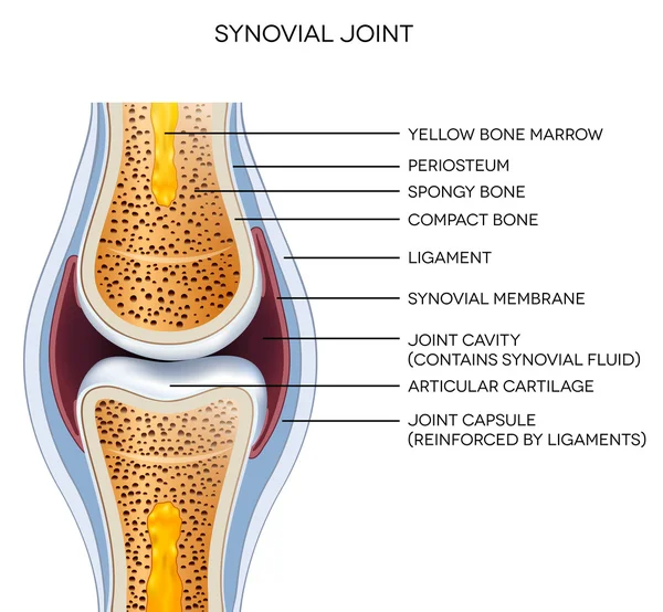
Synovial Joint Chart. Labeled Anatomy Infographic With Two Bones, Articular Cartilage, Joint Cavity, Synovial Fluid, Muscle And Tendon. Isolated Vector Illustration On White.
Vector, 8.73MB, 8055 × 8055 eps
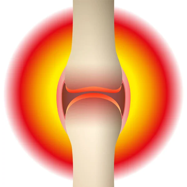
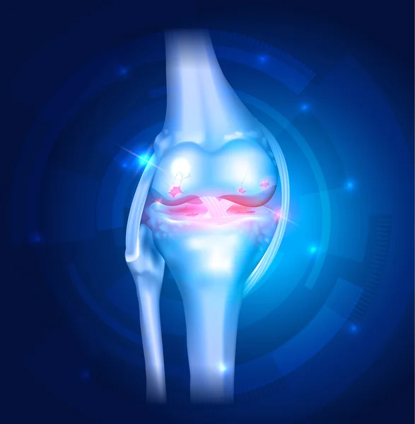
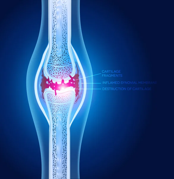
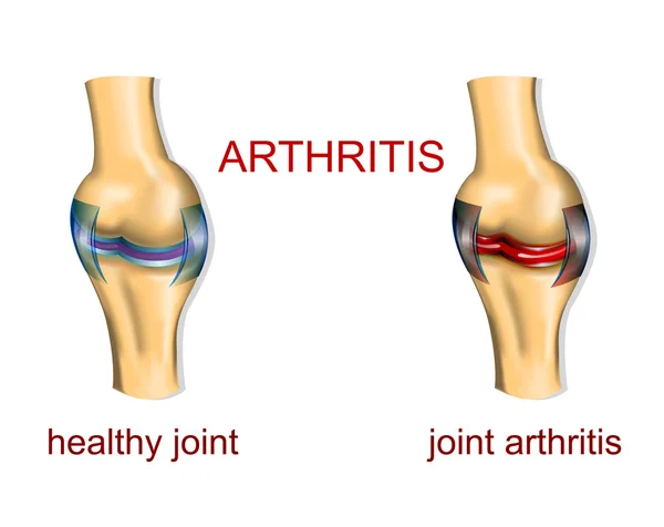
Joint Pain - Schematic Anatomical Graphic Of A Synovial Joint With Arthritis, Rheumatism, Gout, Osteoarthritis Or Inflammation. Isolated Vector Illustration On White Background.
Vector, 2.07MB, 8055 × 8055 eps
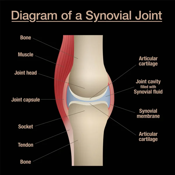
Synovial Joint Diagram. Labeled Anatomy Chart With Two Bones, Articular Cartilage, Joint Cavity, Synovial Fluid, Muscle And Tendon. Isolated Vector Illustration On Black.
Vector, 6.9MB, 8055 × 8055 eps
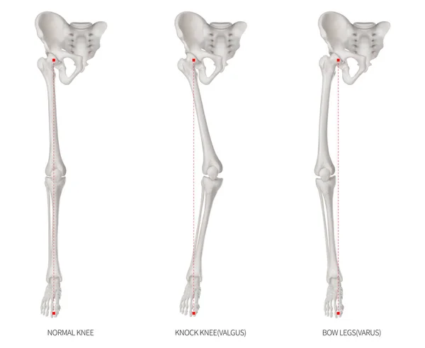
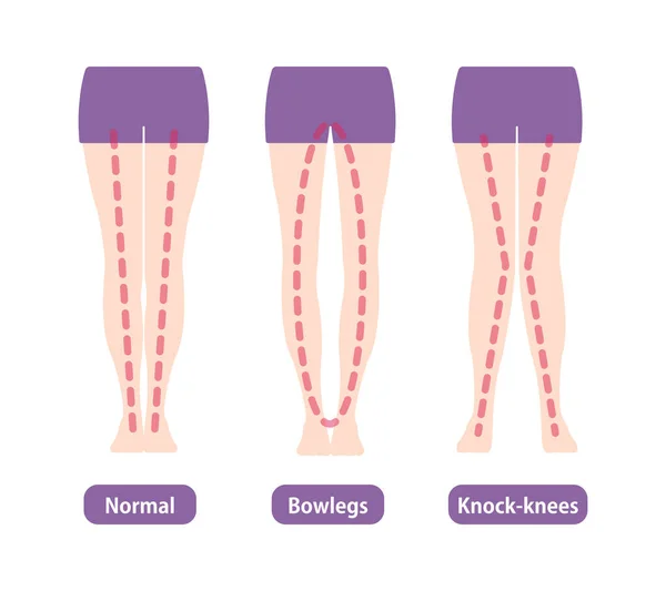
Alignment Types Disease Of Lower Half Limbs Or Leg Bone Problem- Normal- Knock Knee And Bowlegs Or Valgus And Varus Knee- 3D Medical Illustration-human Anatomy And Educational Concept White Background
Image, 6.62MB, 12000 × 9830 jpg
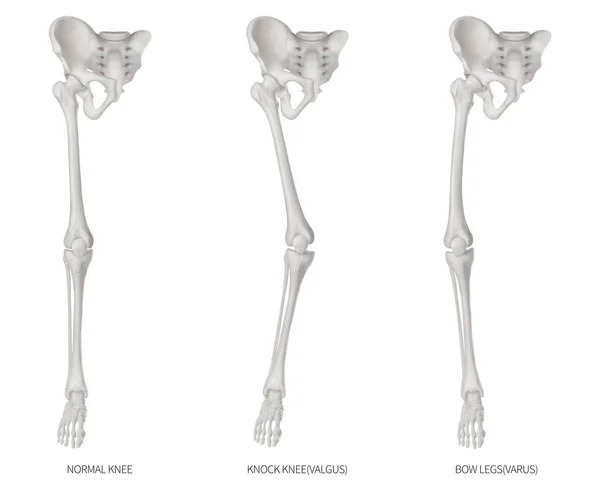
Types Disease Of Lower Half Limbs Or Leg Bone Problem- Normal- Knock Knee And Bowlegs Or Valgus And Varus Knee- 3D Medical Illustration- Human Anatomy And Educational Concept-Isolated White Background
Image, 6.31MB, 12000 × 9830 jpg

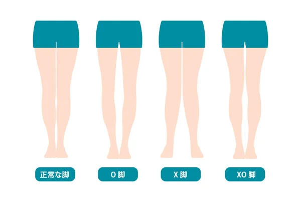
Difference Types Of Legs Angles And Knees Vector Illustration (Japanese)
Vector, 5.59MB, 8088 × 5442 eps
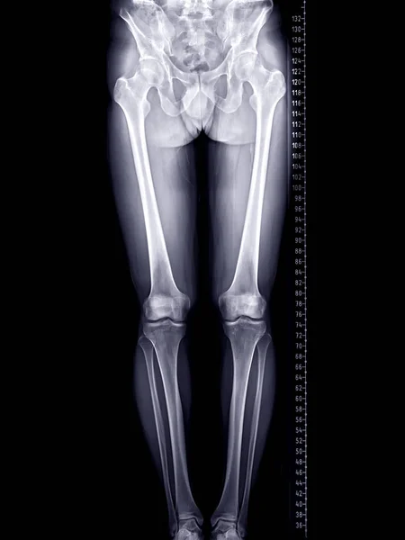
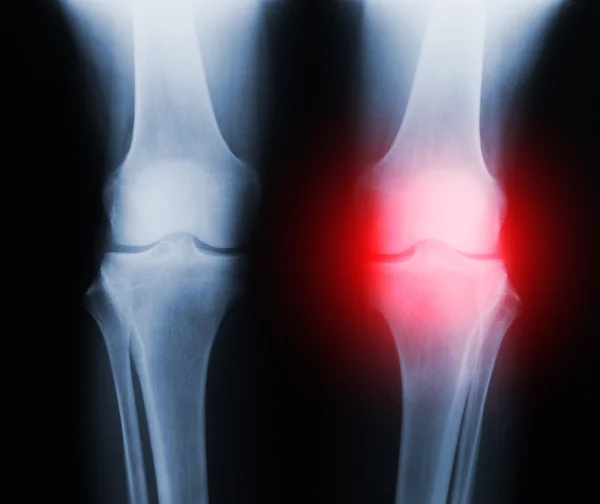
Scanogram Of Lower Limb Or X-ray Image Of Total Lower Extremity With Scale.
Image, 3.15MB, 3280 × 4375 jpg
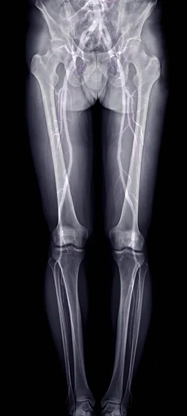
Scanogram Of Lower Limb Or X-ray Image With Merge CTA Femoral Run Off Showing Bone And Vessel Of Lower Limb.
Image, 2.19MB, 1936 × 4303 jpg
Difference Types Of Legs Angles And Knees Vector Illustration (Japanese)
Vector, 5.52MB, 8021 × 5396 eps


The Doctor Gives The Patient An Intra-articular Injection Of A Blockade Into The Elbow Joint. Treatment Of Joints From Arthritis And Osteoarthritis Using Injections With Chondroprotective Agents And Pain Relievers, Vitamins
Image, 1.49MB, 4053 × 2110 jpg
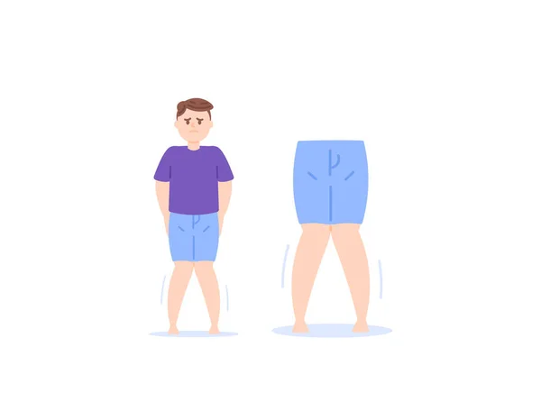
Knock Knee, Rickets, Genu Valgum. Bone Growth Abnormalities. A Man Whose Leg Shape Is X-shaped. Character Illustration Design. Vector Elements
Vector, 0.17MB, 4500 × 3500 ai

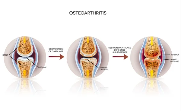


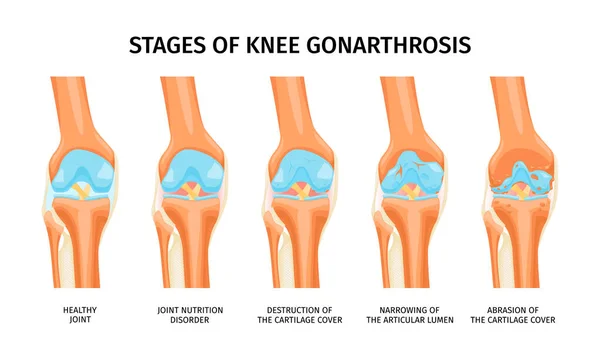
Realistic Infographics Presenting Stages Of Knee Gonarthrosis From Healthy Joint To Abrasion Of Cartilage Cover Vector Illustration
Vector, 0.92MB, 6000 × 3600 eps
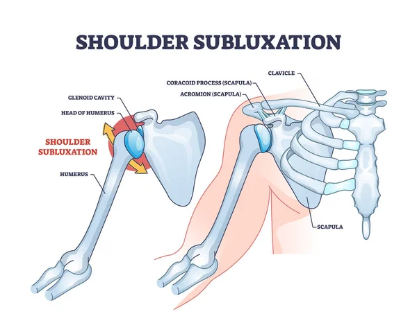
Shoulder Subluxation As Partial Dislocated Arm Joint Problem Outline Diagram. Labeled Educational Medical Scheme With Body Skeletal Anatomy And Dislocated Bones Vector Illustration. Upper Body Trauma.
Vector, 6.41MB, 4600 × 3578 eps


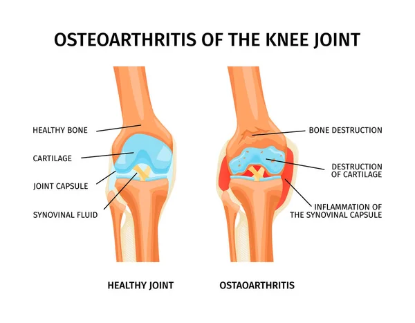
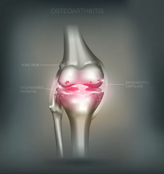
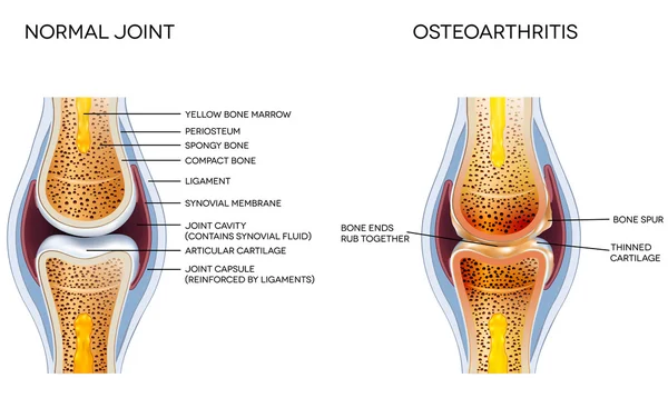
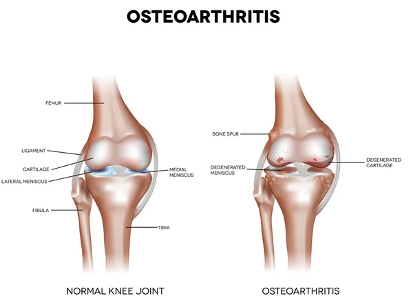
Realistic Infographics With Anatomy Of Healthy Knee And Osteoarthritis Of Joint With Labelled Parts Vector Illustration
Vector, 0.79MB, 4750 × 3701 eps


Realistic Infographics Showing Four Stages Of Rheumatoid Arthritis Of Knee Joint Vector Illustration
Vector, 1.23MB, 6000 × 3600 eps

Osteochondroma Knee Problem As Medical Bone Tumor Overgrowth Outline Diagram
Vector, 6.02MB, 5000 × 3556 eps
Page 1 >> Next