photo free
Home Images Vectors Videos

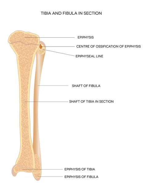
Stock image Joint Cavity page 2

stock vector Abstract colorful joint anatomy icons set

Abstract Colorful Joint Anatomy Icons Set

Vector, 3.28MB, 4800 × 4946 eps
stock vector Osteoporosis

Osteoporosis

Vector, 11.56MB, 5374 × 3579 eps
stock vector Realistic infographics presenting stages of knee gonarthrosis from healthy joint to abrasion of cartilage cover vector illustration

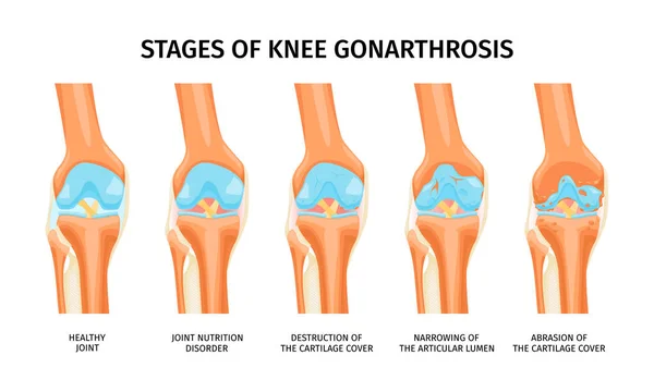
Realistic Infographics Presenting Stages Of Knee Gonarthrosis From Healthy Joint To Abrasion Of Cartilage Cover Vector Illustration

Vector, 0.92MB, 6000 × 3600 eps
stock vector Shoulder subluxation as partial dislocated arm joint problem outline diagram. Labeled educational medical scheme with body skeletal anatomy and dislocated bones vector illustration. Upper body trauma.

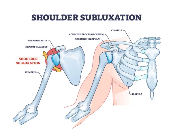
Shoulder Subluxation As Partial Dislocated Arm Joint Problem Outline Diagram. Labeled Educational Medical Scheme With Body Skeletal Anatomy And Dislocated Bones Vector Illustration. Upper Body Trauma.

Vector, 6.41MB, 4600 × 3578 eps
stock vector Hip joint structure

Hip Joint Structure

Vector, 1.47MB, 5000 × 3141 eps
stock image Body

Body

Image, 4.97MB, 5000 × 5000 jpg
stock vector HEALTHY JOINT

HEALTHY JOINT

Vector, 9.25MB, 7288 × 5401 eps
stock vector Realistic infographics with anatomy of healthy knee and osteoarthritis of joint with labelled parts vector illustration

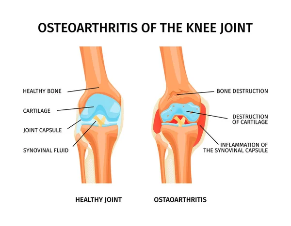
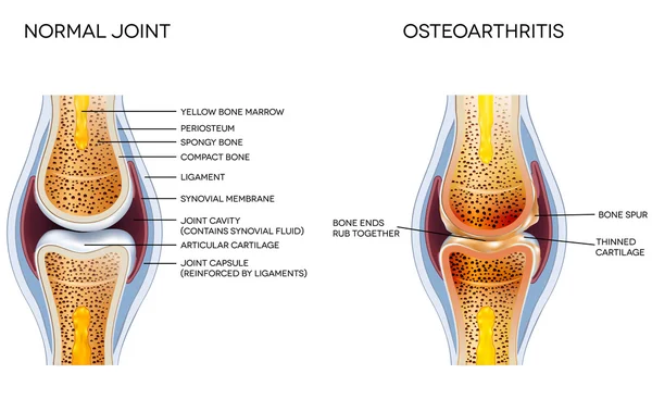
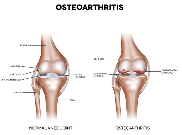
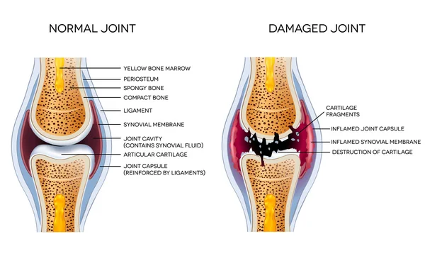
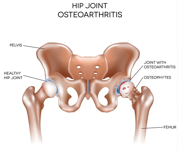
Realistic Infographics With Anatomy Of Healthy Knee And Osteoarthritis Of Joint With Labelled Parts Vector Illustration

Vector, 0.79MB, 4750 × 3701 eps
stock image pelvis bone

Pelvis Bone

Image, 2.07MB, 3072 × 3072 jpg
stock vector orthopedics human anatomy

Orthopedics Human Anatomy

Vector, 1.68MB, 7973 × 7973 eps
stock image Skeleton

Skeleton

Image, 6.57MB, 5000 × 5000 jpg
stock vector Hip joint

Hip Joint

Vector, 9.88MB, 3998 × 5088 eps
stock vector Osteoarthritis and normal joint anatomy

Osteoarthritis And Normal Joint Anatomy

Vector, 7.2MB, 6200 × 3798 eps
stock vector Shoulder joint isolated on white vector

Shoulder Joint Isolated On White Vector

Vector, 2.96MB, 4313 × 4313 eps
stock vector Tibia and Fibula. Leg bones

Tibia And Fibula. Leg Bones

Vector, 1.18MB, 3780 × 4712 eps
stock vector Arthritis of the knee joint

Arthritis Of The Knee Joint

Vector, 14.74MB, 5300 × 4625 eps
stock image 3d render female hip bone

3d Render Female Hip Bone

Image, 3.13MB, 4819 × 3072 jpg
stock image Xray knee joint trauma

Xray Knee Joint Trauma

Image, 2.47MB, 2852 × 2400 jpg
stock vector Knee Osteoarthritis

Knee Osteoarthritis

Vector, 7.42MB, 5700 × 4246 eps
stock image Elbow joint

Elbow Joint

Image, 4.97MB, 4096 × 4096 jpg
stock vector Arthritis of the hip joint

Arthritis Of The Hip Joint

Vector, 1.79MB, 4450 × 5470 eps
stock vector Dislocation of the shoulder joint, medical vector illustration

Dislocation Of The Shoulder Joint, Medical Vector Illustration

Vector, 9.29MB, 5000 × 3000 eps
stock vector Realistic infographics showing four stages of rheumatoid arthritis of knee joint vector illustration

Realistic Infographics Showing Four Stages Of Rheumatoid Arthritis Of Knee Joint Vector Illustration

Vector, 1.23MB, 6000 × 3600 eps
stock vector Osteochondroma knee problem as medical bone tumor overgrowth outline diagram

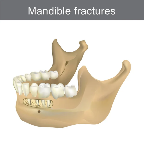
Osteochondroma Knee Problem As Medical Bone Tumor Overgrowth Outline Diagram

Vector, 6.02MB, 5000 × 3556 eps
stock vector Human hip joint and Osteoporosis

Human Hip Joint And Osteoporosis

Vector, 3.77MB, 5000 × 5000 eps
stock vector Female pelvis

Female Pelvis

Vector, 2.21MB, 4296 × 3577 eps
stock vector Shoulder dislocation and humerus bone trauma explanation outline diagram

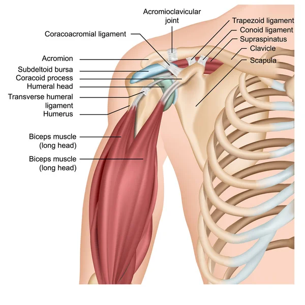
Shoulder Dislocation And Humerus Bone Trauma Explanation Outline Diagram

Vector, 5.87MB, 5000 × 3750 eps
stock vector Knee bursae. synovial pockets or sacs that surround the knee joint cavity. Synovial joint anatomy. Frontal and side view of human knee joint. Vector illustration

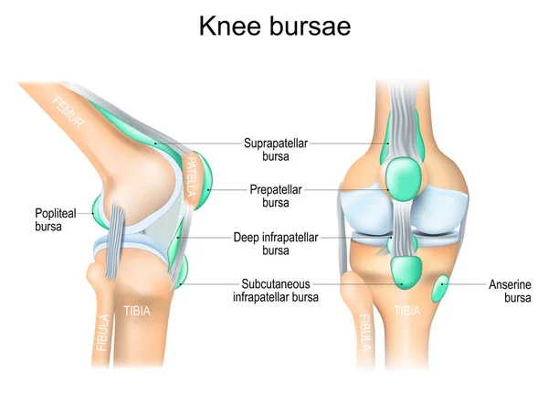
Knee Bursae. Synovial Pockets Or Sacs That Surround The Knee Joint Cavity. Synovial Joint Anatomy. Frontal And Side View Of Human Knee Joint. Vector Illustration

Vector, 6.26MB, 5000 × 3599 eps
stock vector Damaged joint and healthy joint detailed diagram

Damaged Joint And Healthy Joint Detailed Diagram

Vector, 6.67MB, 6200 × 3739 eps
stock vector Male pelvis

Male Pelvis

Vector, 4.19MB, 4178 × 4147 eps
stock image Side View Of Scapula

Side View Of Scapula

Image, 0.47MB, 2877 × 3365 jpg
stock vector Shoulder Joint of Human Body Anatomy infographic diagram with all parts including bones ligaments muscles bursa cavity capsule cartilage membrane for medical science education and health care

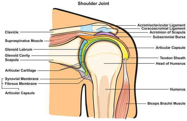
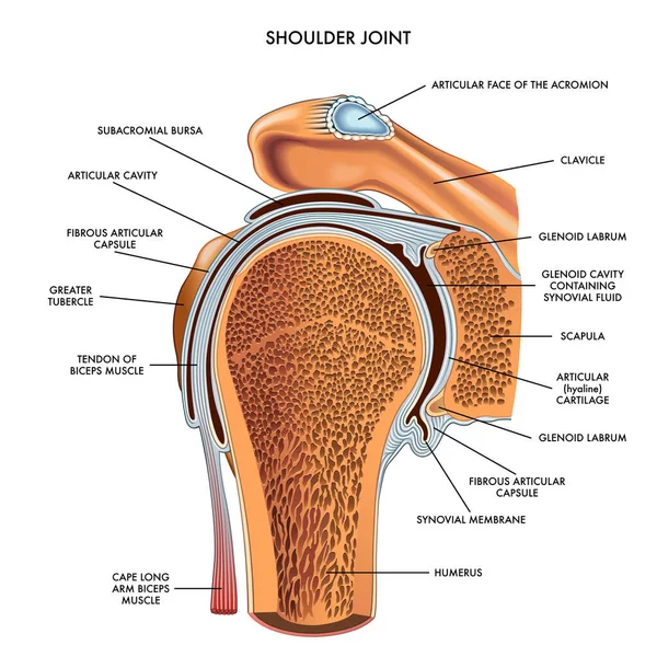
Shoulder Joint Of Human Body Anatomy Infographic Diagram With All Parts Including Bones Ligaments Muscles Bursa Cavity Capsule Cartilage Membrane For Medical Science Education And Health Care

Vector, 0.5MB, 2529 × 1643 eps
stock image 3d render female hip bone

3d Render Female Hip Bone

Image, 2.45MB, 3072 × 3072 jpg
stock vector Osteosarcoma bone tumor as osteogenic sarcoma skeleton cancer outline diagram

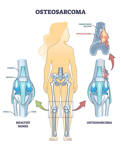
Osteosarcoma Bone Tumor As Osteogenic Sarcoma Skeleton Cancer Outline Diagram

Vector, 6.32MB, 4000 × 4800 eps
stock vector Shoulder anatomy 3d medical vector illustration with arm muscles

Shoulder Anatomy 3d Medical Vector Illustration With Arm Muscles

Vector, 18.12MB, 5000 × 5000 eps
stock image Arthritis of the hip joint on a white background. Illustration

Arthritis Of The Hip Joint On A White Background. Illustration

Image, 1.08MB, 4168 × 4169 jpg
stock image X ray of dog anterior view with Gastric dilatation volvulus GDV or stomach twists- Double bubble pattern indicates stomach torsion has occurred-Veterinary medicine and Veterinary anatomy Concept.

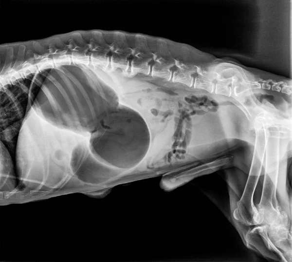
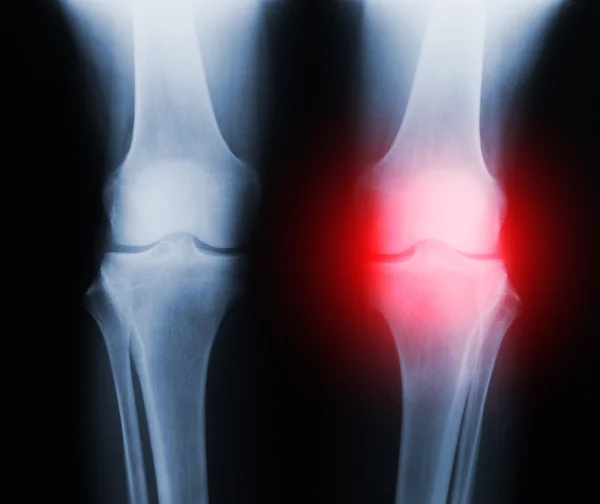
X Ray Of Dog Anterior View With Gastric Dilatation Volvulus GDV Or Stomach Twists- Double Bubble Pattern Indicates Stomach Torsion Has Occurred-Veterinary Medicine And Veterinary Anatomy Concept.

Image, 3.49MB, 5500 × 4958 jpg
stock vector hip bursa anatomy 3d medical vector illustration isolated on white background

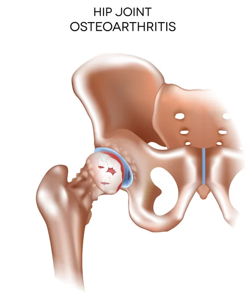
Hip Bursa Anatomy 3d Medical Vector Illustration Isolated On White Background

Vector, 9.2MB, 7000 × 5000 eps
stock vector Osteoarthritis, destruction of cartilage

Osteoarthritis, Destruction Of Cartilage

Vector, 10.64MB, 6100 × 3892 eps
stock vector Arthritis of the hip joint

Arthritis Of The Hip Joint

Vector, 2.51MB, 5350 × 4596 eps
stock vector vector illustration of shoulder joint

Vector Illustration Of Shoulder Joint

Vector, 5.35MB, 4488 × 4488 eps
stock vector Medical problem of osteoporosis disease

Medical Problem Of Osteoporosis Disease

Vector, 0.5MB, 4000 × 4000 eps
stock vector Mandible fractures is the largest, strongest and lowest bone in

Mandible Fractures Is The Largest, Strongest And Lowest Bone In

Vector, 5.33MB, 6500 × 6500 eps
stock vector Healthy bone and broken bone with osteoporosis on the white background.

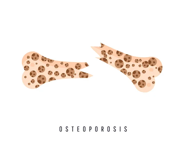
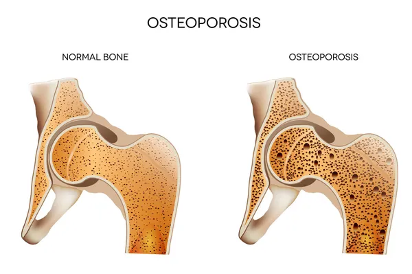
Healthy Bone And Broken Bone With Osteoporosis On The White Background.

Vector, 0.31MB, 4000 × 3200 eps
stock vector Thoracic cage is made up of bones and cartilage along, It consists of the 12 pairs of ribs with their costal cartilages and the sternum. Illustration human bones.

Thoracic Cage Is Made Up Of Bones And Cartilage Along, It Consists Of The 12 Pairs Of Ribs With Their Costal Cartilages And The Sternum. Illustration Human Bones.

Vector, 10.99MB, 7000 × 7000 eps
stock vector Rheumatism medical health care vector illustration poster diagram with damaged knee erosion and painful body joints.

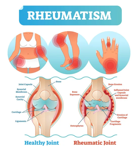
Rheumatism Medical Health Care Vector Illustration Poster Diagram With Damaged Knee Erosion And Painful Body Joints.

Vector, 5.64MB, 4228 × 4639 eps
stock image pelvis bone

Pelvis Bone

Image, 1.67MB, 3072 × 3072 jpg
stock vector The Thoracic Cage. The thoracic cage is made up of bones and cartilage along, It consists of the 12 pairs of ribs with their costal cartilages and the sternum.

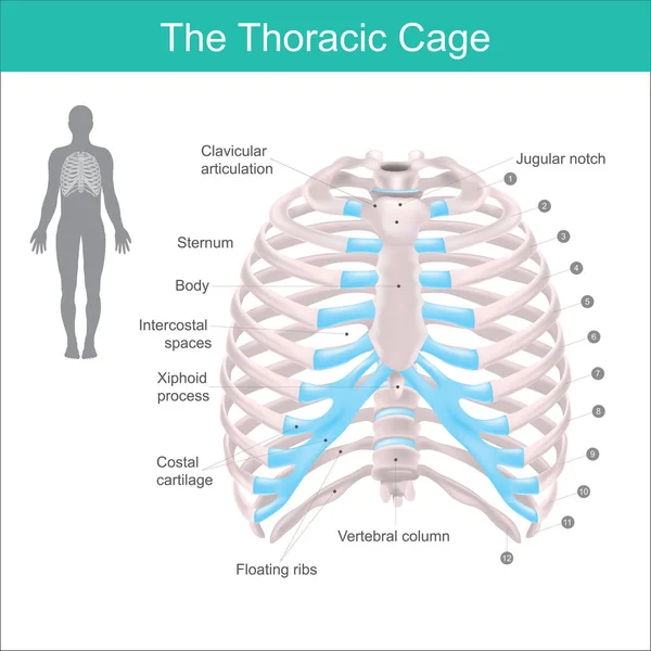
The Thoracic Cage. The Thoracic Cage Is Made Up Of Bones And Cartilage Along, It Consists Of The 12 Pairs Of Ribs With Their Costal Cartilages And The Sternum.

Vector, 11.17MB, 5266 × 5266 eps
stock image Articular capsule Anatomy For Medical Concept 3D Illustration

Articular Capsule Anatomy For Medical Concept 3D Illustration

Image, 4.91MB, 4096 × 4096 jpg
stock image Structure of bone

Structure Of Bone

Image, 4.33MB, 5400 × 4096 jpg
Previous << Page 2 >> Next