
Stock image Kinase page 2
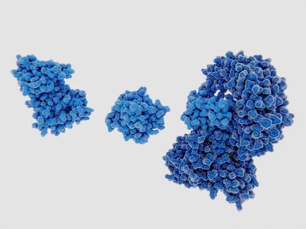
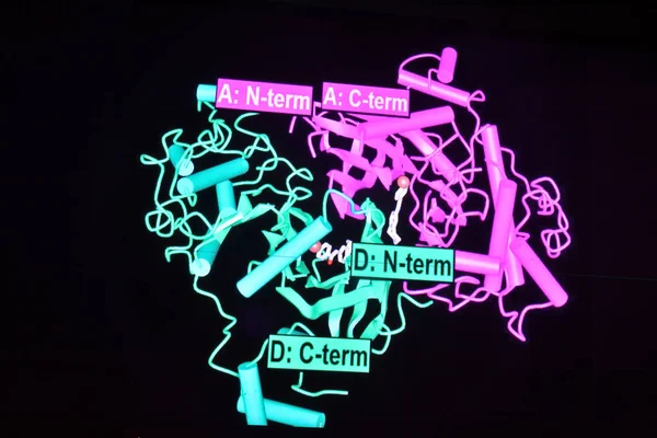
The Mitotic Kinase NEK7 Binds To Inactive NRLP3 Leading To The Activation Of The Inflammasome
Image, 3.59MB, 8000 × 6000 jpg
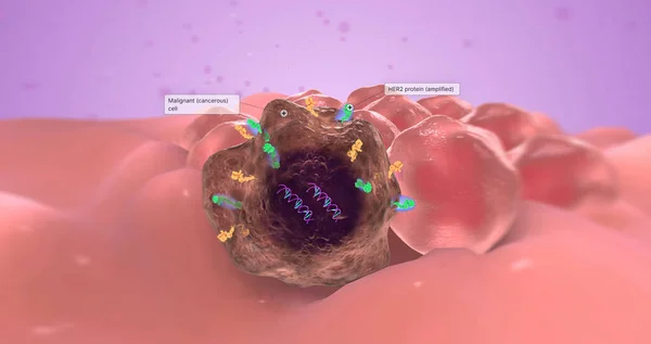
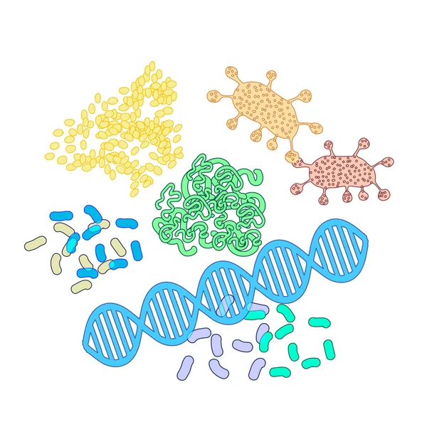
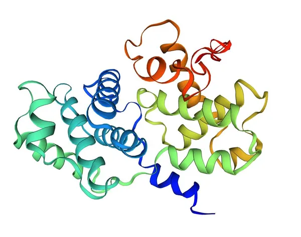
The Abnormal Gene Is Known As An Oncogene Because It Causes Tumor Growth. 3D Rendering
Image, 2.86MB, 7340 × 3884 jpg
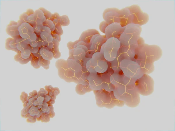
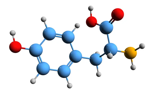
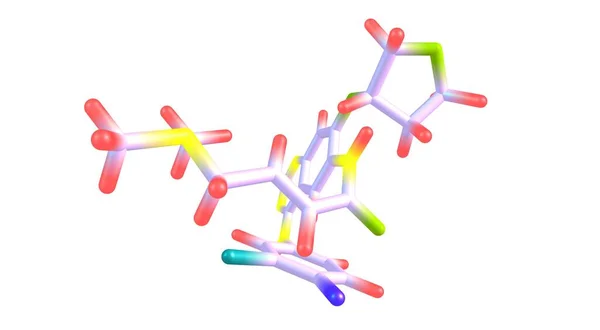
3D Image Of Tyrosine Skeletal Formula - Molecular Chemical Structure Of 4-hydroxyphenylalanine Isolated On White Background
Image, 2.01MB, 5500 × 3630 jpg
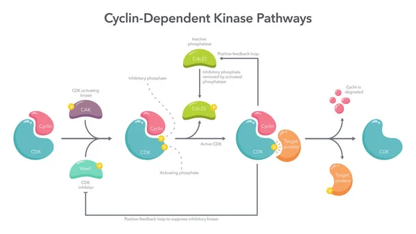
Cyclin Dependent Kinase Activation Pathway Science Vector Illustration Infographic
Vector, 0.45MB, 8333 × 4688 ai
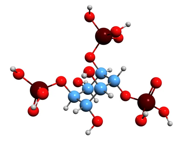
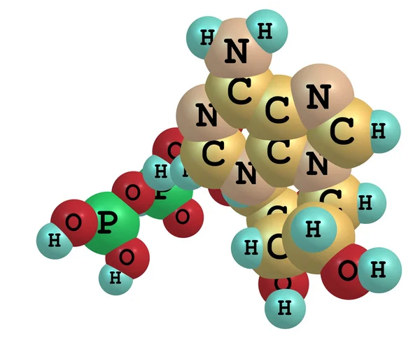
3D Image Of Inositol Trisphosphate Skeletal Formula - Molecular Chemical Structure Of Inositol Phosphate Signaling Molecule Isolated On White Background
Image, 3.87MB, 6943 × 5520 jpg

Hand Writing Creatine With Blue Marker On Transparent Wipe Board Isolated On White Background.
Image, 2.69MB, 5616 × 3744 jpg
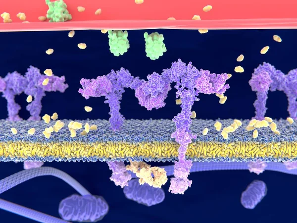
Insulin (green) Binding To The Insulin Receptor (violet) Activates The Transport Of Glucose (yellow) Into The Cell. Illustration
Image, 3.71MB, 8000 × 6000 jpg

Structure Of Cyclin-dependent Kinase 2 (CDK2, Blue) In Complex With Cyclin E (brown). 3D Cartoon And Gaussian Surface Models, PDB 1w98, White Background
Image, 3.84MB, 8000 × 4000 jpg

Insulin (green) Binding To The Insulin Receptor (violet) Activates The Transport Of Glucose (yellow) Into The Cell (phase 1). Illustration
Image, 3.96MB, 8000 × 6000 jpg

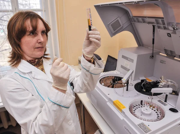
Doctor Holding A Test Blood Sample Tube With EGFR Test On The Background Of Medical Test Tubes With Analyzes.
Image, 8.65MB, 5789 × 3859 jpg

Alectinib Molecule Is An Oral Drug That Blocks The Activity Of Anaplastic Lymphoma Kinase And Is Used To Treat Non-small-cell Lung Cancer. 3d Illustration
Image, 0.73MB, 7000 × 3638 jpg
Enzymology Typography, Wordart, Wordcloud, Enzyme, Enzymology, Science, Chemical
Image, 0.92MB, 6400 × 4800 jpg

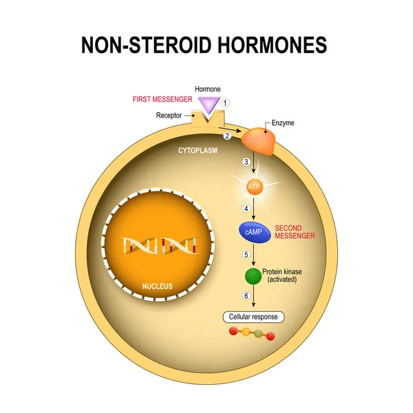
Difference Between Non-steroid Hormones And Steroid Hormones. Steroid Hormones Pass Through The Cell Membrane. Inside The Cell They Bind To Specific Receptors, Influencing Gene Expression And Protein Synthesis. Non-steroidal Hormones Bind To Receptor
Vector, 11.97MB, 4444 × 4444 eps

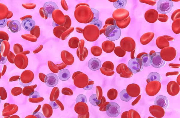
High Level Of RBC And Platelet In Polycythemia Vera (PV) - Closeup View 3d Illustration
Image, 5.49MB, 10000 × 6600 jpg

Doctor Holding A Test Blood Sample Tube With EGFR Test On The Background Of Medical Test Tubes With Analyzes.
Image, 9.04MB, 6000 × 4000 jpg

Thymoma Icon. Trendy Flat Vector Thymoma Icon On White Background From Diseases Collection, Vector Illustration Can Be Use For Web And Mobile, Eps10
Vector, 0.58MB, 6944 × 6944 eps
Thymoma Icon. Trendy Modern Flat Linear Vector Thymoma Icon On White Background From Thin Line Diseases Collection, Editable Outline Stroke Vector Illustration
Vector, 0.6MB, 6944 × 6944 eps
Afatinib Is A Medication Used To Treat Non-small Cell Lung Carcinoma, NSCLC. 3d Illustration
Image, 0.69MB, 7000 × 3855 jpg

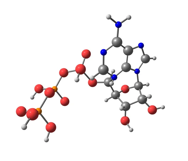
AICA Ribonucleotide, AICAR Molecule. It Is Aminoimidazole, Cardiovascular Drug, Plant And Human Metabolite. Structural Chemical Formula On The Dark Blue Background. Vector Illustration
Vector, 5.26MB, 5000 × 5000 eps
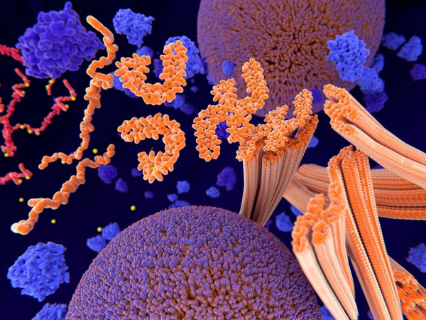
Pathological Phosphorylation (yellow) Of Tau Proteins (red-orange) Leads To Disintegration Of Microtubuli In The Neuron Axon An Aggregation Of The Tau Proteins. The Transport Of Synaptic Vesicles (orange-violet Spheres) Is Interrupted.
Image, 9.48MB, 8000 × 6000 jpg
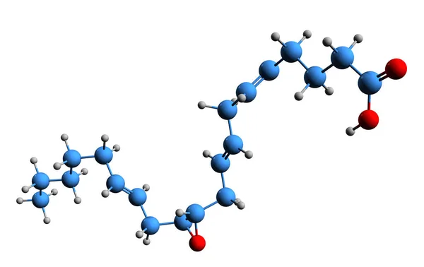
3D Image Of 11,12-Epoxyeicosatrienoic Acid Skeletal Formula - Molecular Chemical Structure Of 11,12-Eet Isolated On White Background
Image, 1.59MB, 5000 × 3156 jpg

Heat-shock Protein 90 Dimer (pink-blue)-HSP90 Co-chaperone Cdc37 (yellow)-cyclin-dependent Kinase 4 (green) Complex. 3D Cartoon And Gaussian Surface Models, PDB 5fwk, White Background
Image, 3.93MB, 8000 × 3000 jpg

High Level Of RBC And Platelet In Polycythemia Vera (PV) - Isometric View 3d Illustration
Image, 7.68MB, 10000 × 6600 jpg


Cardiac And Cardiovascular Biomarkers - Medical Innovation And Advances In Cardiovascular Biomarker Discovery - Conceptual Illustration
Image, 11.83MB, 9584 × 5298 jpg


Systematic Observation - Learn, Study And Inspect It. Taking A Closer Look At Systematic Observation.
Image, 2.71MB, 5472 × 3648 jpg

Alectinib Molecule Is An Oral Drug That Blocks The Activity Of Anaplastic Lymphoma Kinase And Is Used To Treat Non-small-cell Lung Cancer. 3d Illustration
Image, 1MB, 7000 × 3638 jpg

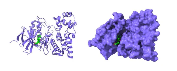
Crystal Structure Of Human JNK3 Complexed With An Isoquinolone Inhibitor. 3D Cartoon And Gaussian Surface Models, PDB 2zdu, White Background
Image, 4.77MB, 10000 × 4000 jpg
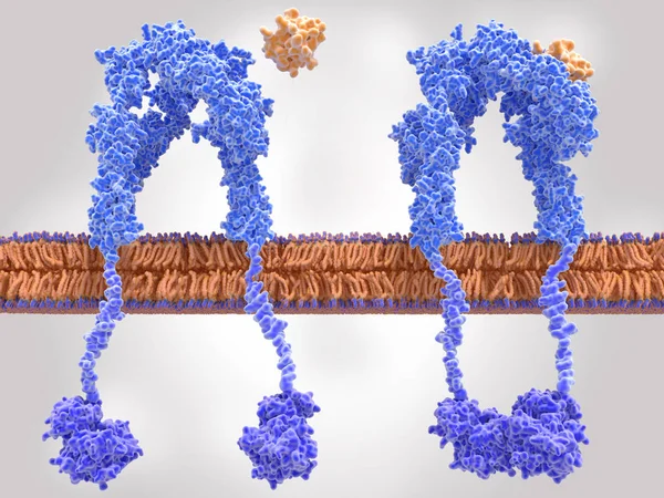
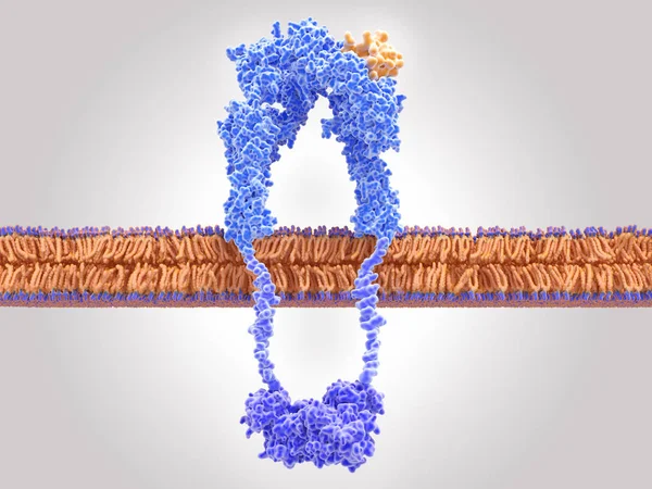
Insulin Receptor Inactivated (left) And Activated (right) After Insulin Binding
Image, 9MB, 8000 × 6000 jpg
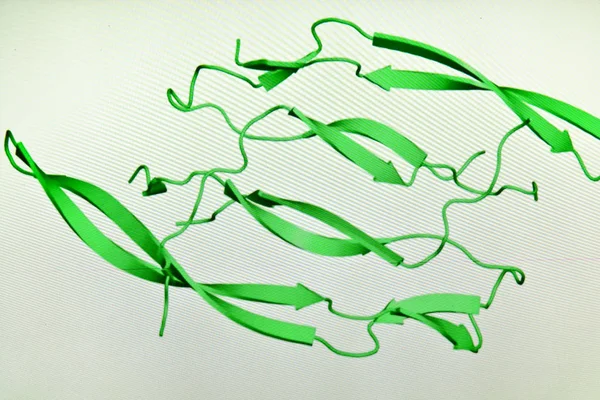
Crystal Structure Of Human Cyclin B1. 3D Cartoon Model, PDB 2b9r, White Background
Image, 1.81MB, 7850 × 6190 jpg


High Level Of Red Blood Cell In Polycythemia Vera (PV) - Closeup View 3d Illustration
Image, 6.44MB, 10000 × 6600 jpg
High Level Of Red Blood Cell In Polycythemia Vera (PV) - Isometric View 3d Illustration
Image, 7.99MB, 10000 × 6600 jpg
Previous << Page 2 >> Next