
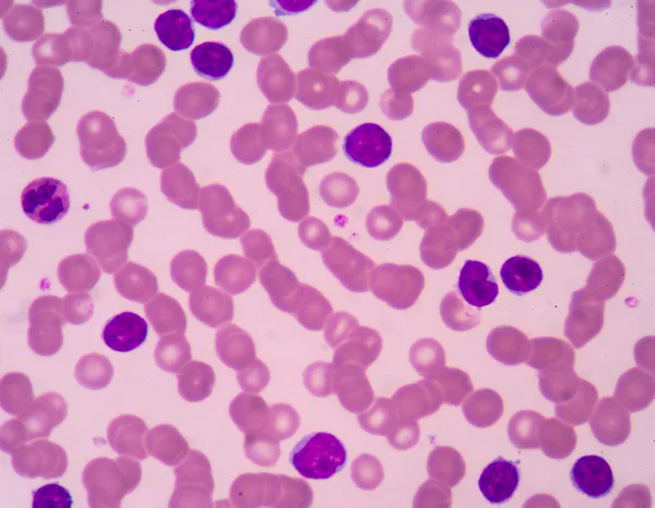
Stock image Lymphocyte page 4


Vaccination Campaign Promoting Immunization With Happy Characters: Winking Syringe, Vaccine Vial, Smiling Lymphocytes And Shield Decorated With Cross.
Vector, 1.78MB, 5000 × 4429 eps


Eosinophilic Cationic Protein Is Incorporated Into The Membranes Of Helminth Cells And Disrupts Their Integrity. Eosinophil Is A Blood Cell. Vector Illustration On Isolated Background
Vector, 16.6MB, 5000 × 5000 eps

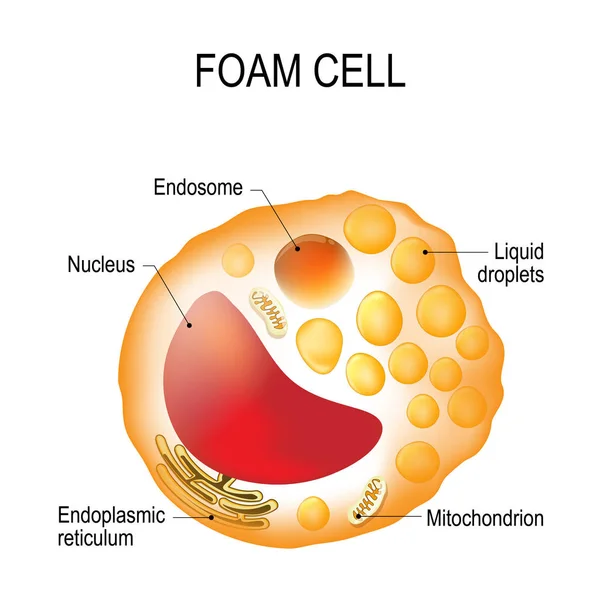
The Structure Of Platelets. Platelets Are A Blood Cell. Myeloid, Stem, Cell, Megakaryocyte, Megakaryoblast. Infographics. Illustration On Isolated Background.
Image, 1.36MB, 5208 × 5208 jpg

Diagnosis Of Lymphocytopenia. Decreased Lymphocytes In The Blood. Infographics. Vector Illustration On Isolated Background.
Vector, 33.73MB, 5000 × 4868 eps

Symptoms Of Lymphocytopenia. Decreased Lymphocytes In The Blood. Infographics. Vector Illustration On Isolated Background.
Vector, 33.17MB, 5000 × 4868 eps


Ponesimod, Experimental Anti-inflammatory Drug Molecule. Treatment Of Multiple Sclerosis MS And Psoriasis. Structural Chemical Formula And Molecule Model. Vector Illustration
Vector, 5.29MB, 5000 × 5000 eps


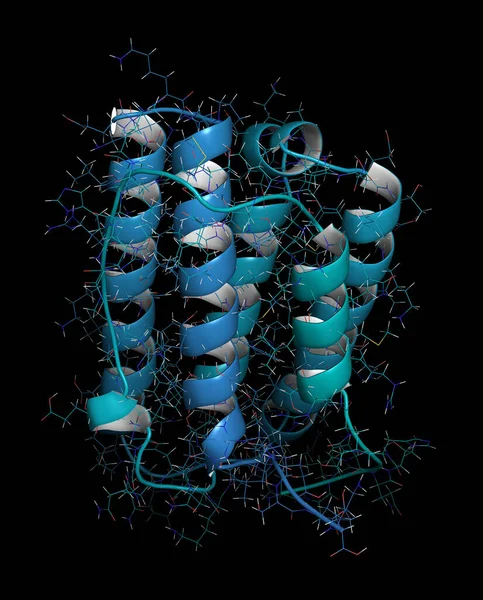
Interferon Alpha 2a (IFNA2) Molecule, 3D Rendering. Pegylated Analogs Of This Cytokine Are Used To Treat Hepatitis B And C Infections.
Image, 11.43MB, 6450 × 8000 jpg


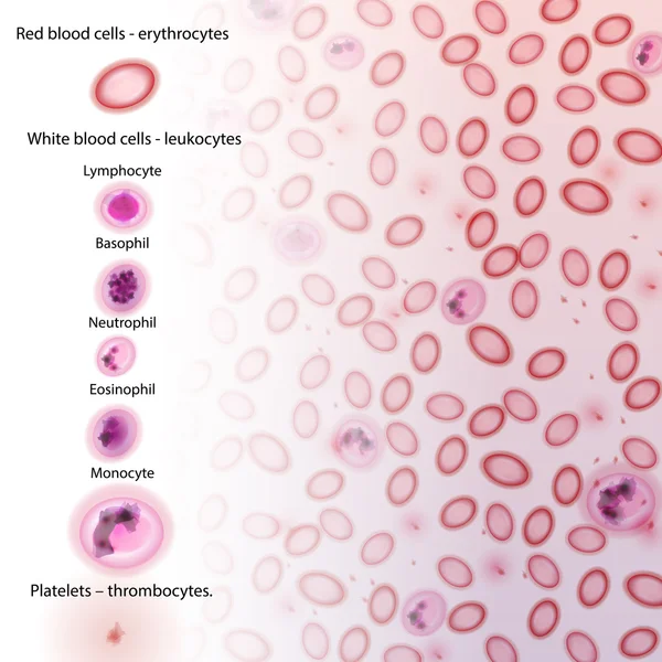
Lymphatic System Isolated Anatomical Structure Medicine And Healthcare
Vector, 1.17MB, 5058 × 7244 eps
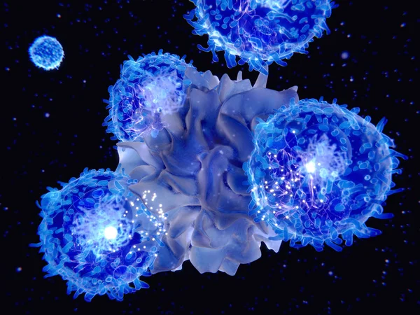
3d Computer Illustration Of A Dendritic Cell. They Areantigen-presenting Cells Of The Immune System. Their Main Function Is To Process Antigen Material And Present It On The Cell Surface To The T Cells Of The Immune System. They Are Messengers Betwe
Image, 5.2MB, 8000 × 6000 jpg
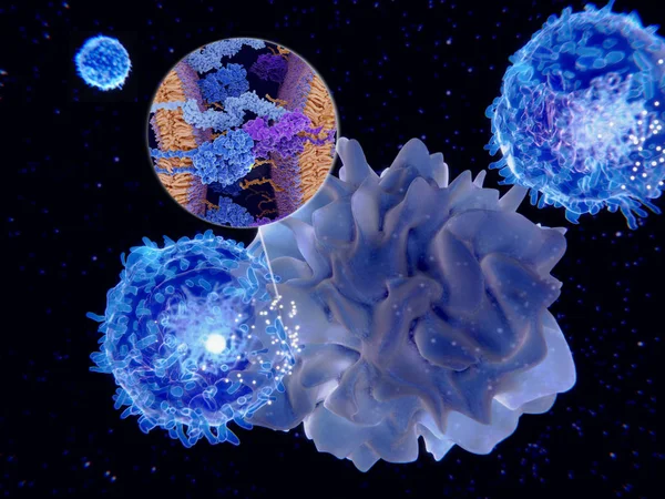
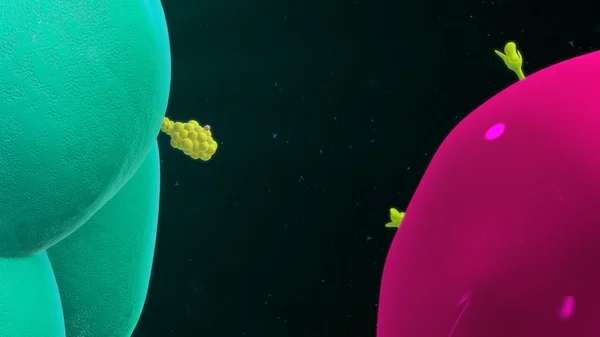
Dendritic Cells Present Antigens (green) To Lymphocytes Through Their Membran Bound MHC-molecules (violet). CD4 Molecules (light Blue) Bind To Other Portions Of The MHC, Strengthening The Interaction.
Image, 10.24MB, 8000 × 6000 jpg
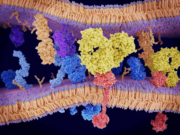
Cancer Cells Express PD-L1 (orange) Proteins On Their Surface To Trick The Immune System. The Interaction Of PD-L1 With PD-1 Of T-cells Leads To A Down-regulation Of T-cells. The Antibody (yellow) Blocks This Interaction.
Image, 18.3MB, 8000 × 6000 jpg
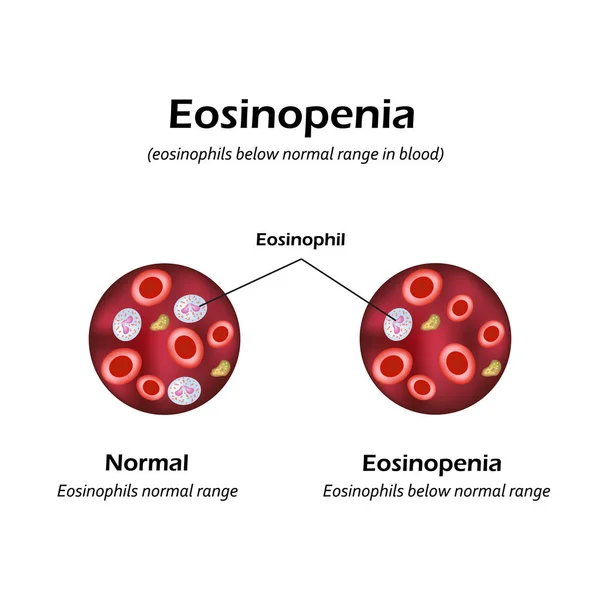
Eosinophils Below Normal Range In Blood. Eosinopenia. Infographics. Vector Illustration
Vector, 6.45MB, 5000 × 5000 eps


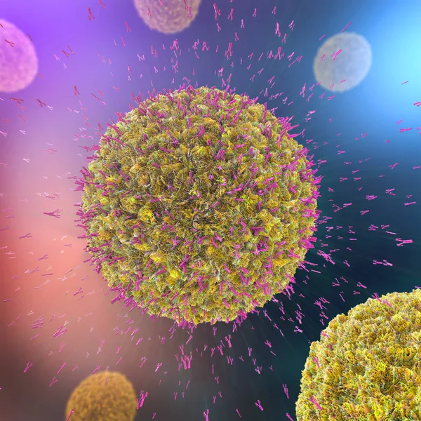
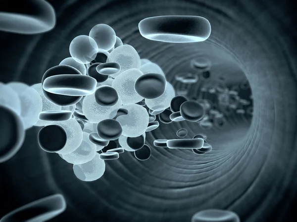
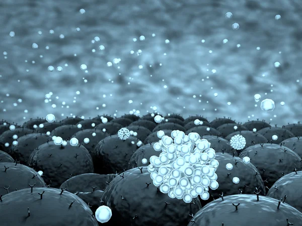
3d Illustration Of Antibodies Attacking Virus Cell Into The Bloodstream
Image, 26.94MB, 8000 × 8000 jpg

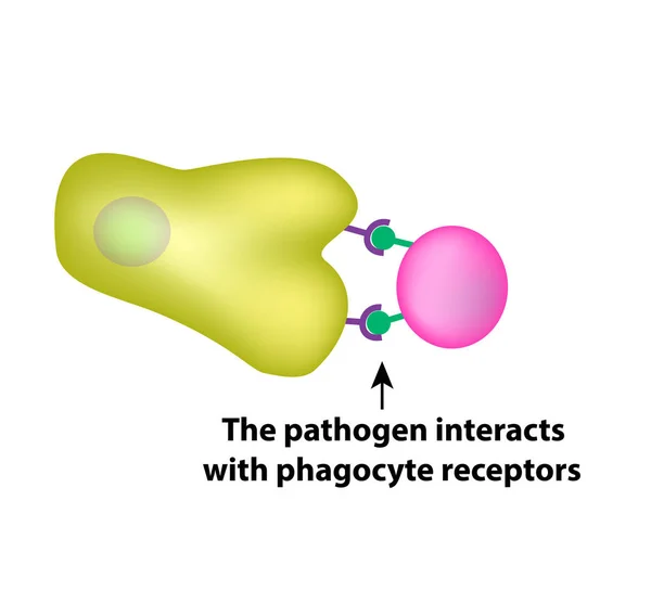
Innate Immunity. Adaptive Specific . Phagocytosis. Infographics. Vector Illustration
Vector, 1.45MB, 5000 × 4734 eps
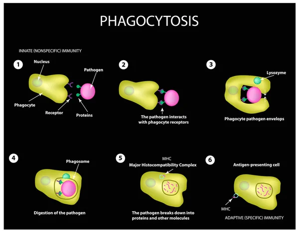
Innate Immunity. Adaptive Specific . Phagocytosis. Infographics. Vector
Vector, 3.99MB, 5000 × 3911 eps

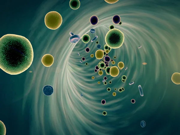
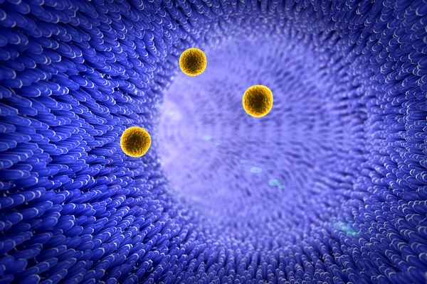
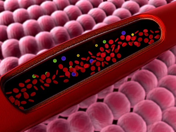
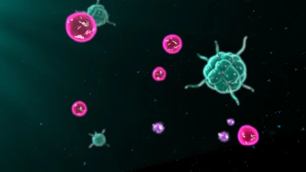
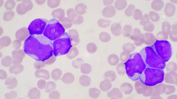
Primary Myelofibrosis (PMF) Cells In Blood Flow - Closeup View 3d Illustration
Image, 6.32MB, 10000 × 6600 jpg
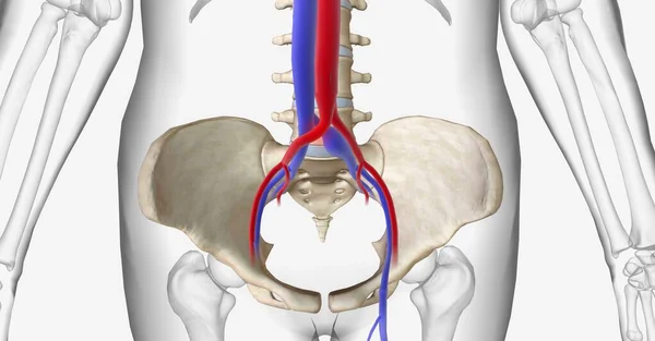
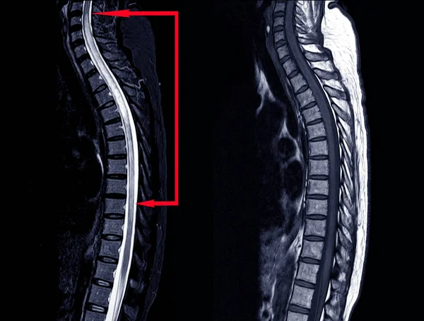
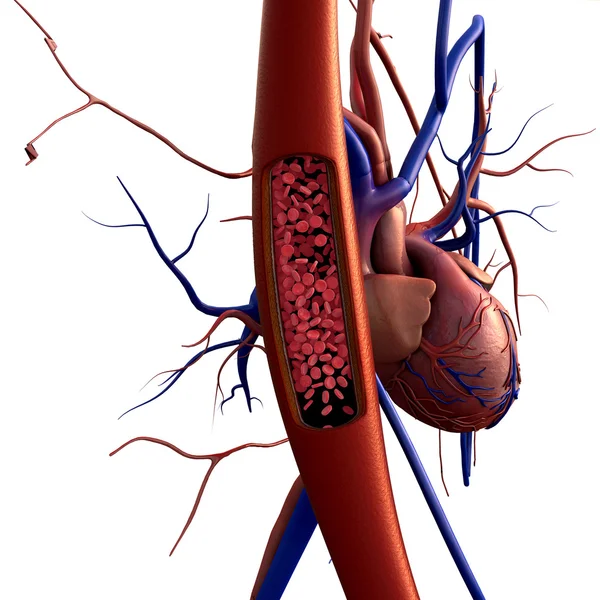
May Thurner Syndrome Is Compression Of The Left Common Iliac Vein Between The Right Common Iliac Artery And The 5th Lumbar Vertebra Of The Spine. 3D Rendering
Image, 16.81MB, 11115 × 5802 jpg

Previous << Page 4 >> Next