photo free
Home Images Vectors Videos

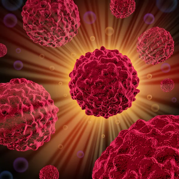
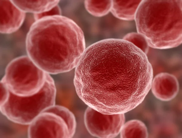
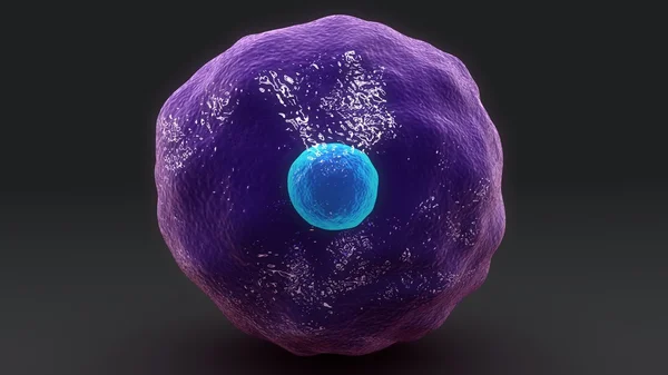
Stock image Malignant Cell

stock vector Tumor cells concept

Tumor Cells Concept

Vector, 0.91MB, 4168 × 4168 eps
stock vector Cancer cell and normal cell. Healthy tissue and Malignant tumour. illustration showing cancer disease development. Vector diagram for your design, educational, biological, science and medical use

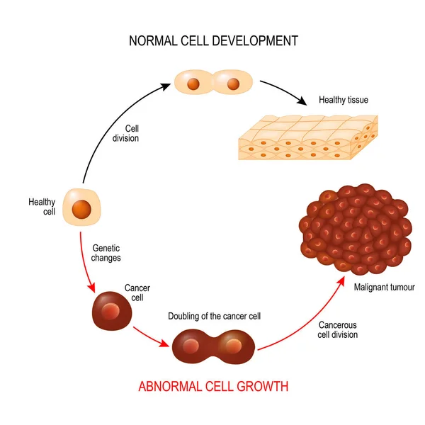
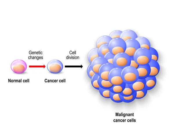
Cancer Cell And Normal Cell. Healthy Tissue And Malignant Tumour. Illustration Showing Cancer Disease Development. Vector Diagram For Your Design, Educational, Biological, Science And Medical Use

Vector, 2.66MB, 6102 × 6102 eps
stock vector Cancer cells. illustration showing cancer disease development. Healthy tissue and Malignant tumour.  Vector diagram for your design, educational, biological, science and medical use

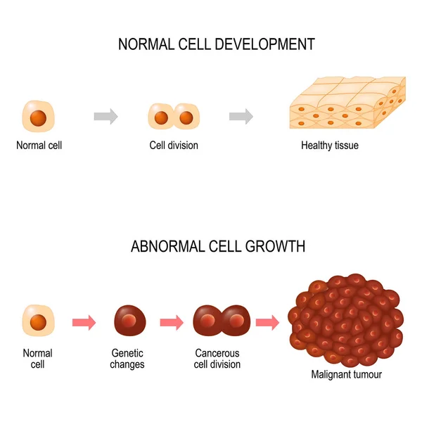
Cancer Cells. Illustration Showing Cancer Disease Development. Healthy Tissue And Malignant Tumour. Vector Diagram For Your Design, Educational, Biological, Science And Medical Use

Vector, 2.5MB, 5207 × 5206 eps
stock image 3d computer illustration of a dendritic cell. They areantigen-presenting cells  of the immune system. Their main function is to process antigen material and present it on the cell surface to the T cells of the immune system. They are messengers betwe

3d Computer Illustration Of A Dendritic Cell. They Areantigen-presenting Cells Of The Immune System. Their Main Function Is To Process Antigen Material And Present It On The Cell Surface To The T Cells Of The Immune System. They Are Messengers Betwe

Image, 5.2MB, 8000 × 6000 jpg
stock image Dendritic cells present antigens (green) to lymphocytes through their membran bound MHC-molecules (violet). CD4 molecules (light blue) bind to other portions of the MHC, strengthening the interaction.

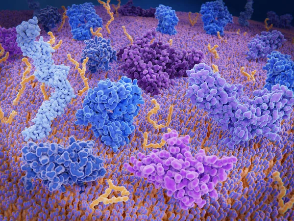
Dendritic Cells Present Antigens (green) To Lymphocytes Through Their Membran Bound MHC-molecules (violet). CD4 Molecules (light Blue) Bind To Other Portions Of The MHC, Strengthening The Interaction.

Image, 10.24MB, 8000 × 6000 jpg
stock vector Normal human cell, cancer cell and malignant cancer

Normal Human Cell, Cancer Cell And Malignant Cancer

Vector, 2.69MB, 7232 × 5492 eps
stock vector Normal cells, hypertrophy is an increase in cell size, hyperplasia results from an increase in cell number. different. Vector diagram for educational, medical, biological and science use

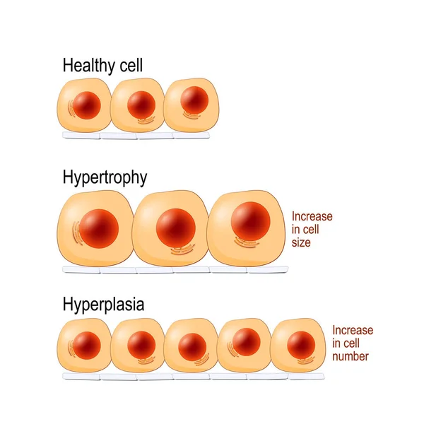
Normal Cells, Hypertrophy Is An Increase In Cell Size, Hyperplasia Results From An Increase In Cell Number. Different. Vector Diagram For Educational, Medical, Biological And Science Use

Vector, 7.14MB, 4462 × 4462 eps
stock image Interactions of MHC-II with the T-cell receptor and CD4  and B7-1 with CD-28 activates T-cells while the interactions of P7-1 with CTLA-4  and PD-L1  with PD-1 deactivates T-cells.

Interactions Of MHC-II With The T-cell Receptor And CD4 And B7-1 With CD-28 Activates T-cells While The Interactions Of P7-1 With CTLA-4 And PD-L1 With PD-1 Deactivates T-cells.

Image, 10.7MB, 8000 × 6000 jpg
stock image PD-1  (red) extends from the surface of a T-cell and interacts with the ligand protein PD-L1 (yellow) from a antigen presenting cell. Although the T-cell has been activated through the interaction of  a T-cell receptor (blue) and a MHC protein (viole

PD-1 (red) Extends From The Surface Of A T-cell And Interacts With The Ligand Protein PD-L1 (yellow) From A Antigen Presenting Cell. Although The T-cell Has Been Activated Through The Interaction Of A T-cell Receptor (blue) And A MHC Protein (viole

Image, 18.32MB, 8000 × 6000 jpg
stock image Interaction of MHC-II (red) with the T-cell receptor (blue) and CD4 (light blue) and B7-1 (orange) with CD-28 (dark blue) activates T-cells while the interaction of P7-1 with CTLA-4 (violett) and PD-L1 (yellow) with PD-1 deactivates T-cells

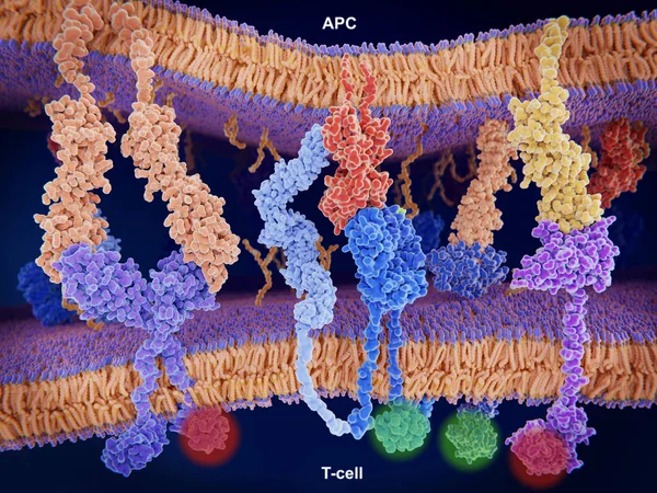
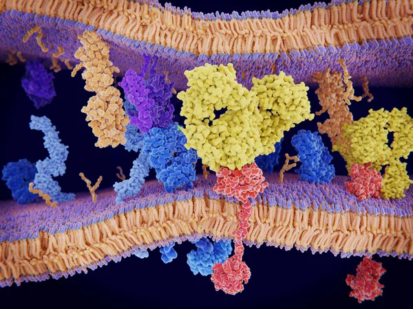
Interaction Of MHC-II (red) With The T-cell Receptor (blue) And CD4 (light Blue) And B7-1 (orange) With CD-28 (dark Blue) Activates T-cells While The Interaction Of P7-1 With CTLA-4 (violett) And PD-L1 (yellow) With PD-1 Deactivates T-cells

Image, 10.65MB, 8000 × 6000 jpg
stock image Cancer cells express PD-L1 (orange) proteins on their surface to trick the immune system. The interaction of PD-L1 with PD-1 of  T-cells leads to a down-regulation of T-cells. The antibody (yellow) blocks this interaction.

Cancer Cells Express PD-L1 (orange) Proteins On Their Surface To Trick The Immune System. The Interaction Of PD-L1 With PD-1 Of T-cells Leads To A Down-regulation Of T-cells. The Antibody (yellow) Blocks This Interaction.

Image, 18.3MB, 8000 × 6000 jpg
stock image Viruses Cells Killing Cancer as an Oncolytic Virus immunology and immunotherapy Therapy to kill cancers by attacking the malignant tumor cell and infecting them and destroying the pathogen as a 3D render.

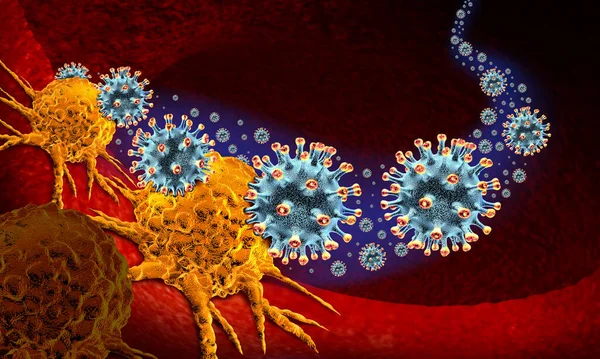
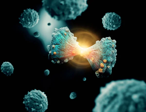
Viruses Cells Killing Cancer As An Oncolytic Virus Immunology And Immunotherapy Therapy To Kill Cancers By Attacking The Malignant Tumor Cell And Infecting Them And Destroying The Pathogen As A 3D Render.

Image, 11.48MB, 6685 × 4008 jpg
stock image Immunologically active proteins on a T-cell. TCR (blue), CD-4 (light blue), CD-28 (dark blue),  PD-1 (magenta), CTLA-4 (violet), Ca-channel (dark violet). The T-cell receptor, CD-4 and CD-28 activate T-cells, while PD-1 and CTLA-4 inhibit the activat

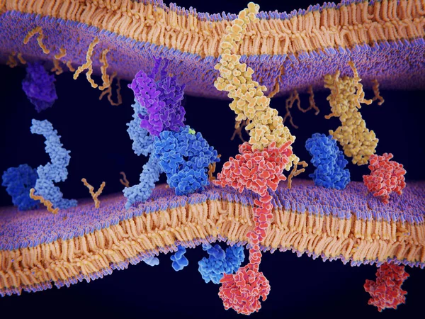
Immunologically Active Proteins On A T-cell. TCR (blue), CD-4 (light Blue), CD-28 (dark Blue), PD-1 (magenta), CTLA-4 (violet), Ca-channel (dark Violet). The T-cell Receptor, CD-4 And CD-28 Activate T-cells, While PD-1 And CTLA-4 Inhibit The Activat

Image, 10.2MB, 8000 × 6000 jpg
stock image Oncolytic Virus immunotherapy and cancer vaccine Therapy as a treatment to kill cancers by attacking the malignant tumor cell and infecting them and destroying the pathogen as a 3D render.

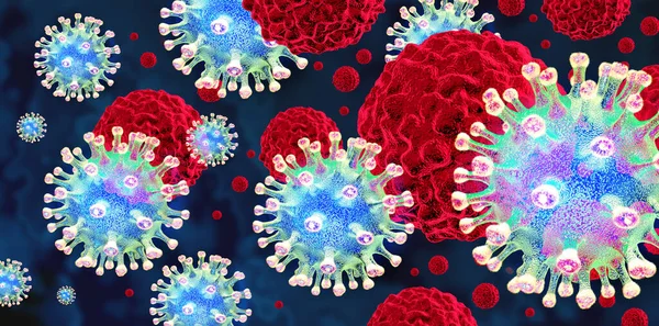
Oncolytic Virus Immunotherapy And Cancer Vaccine Therapy As A Treatment To Kill Cancers By Attacking The Malignant Tumor Cell And Infecting Them And Destroying The Pathogen As A 3D Render.

Image, 15.49MB, 7477 × 3700 jpg
stock image Medicine Resistant Cancer as a deadly Carcinoma mutated viral cell attacking a pharmaceutical pill with a punch as a medical pathology and oncology disease risk as a 3D illustration

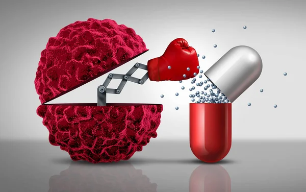
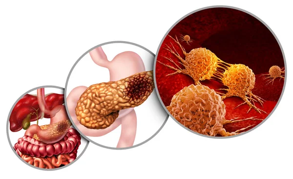
Medicine Resistant Cancer As A Deadly Carcinoma Mutated Viral Cell Attacking A Pharmaceutical Pill With A Punch As A Medical Pathology And Oncology Disease Risk As A 3D Illustration

Image, 7.39MB, 6208 × 3900 jpg
stock image Cancer cells

Cancer Cells

Image, 5.25MB, 2945 × 2945 jpg
stock vector 3D Isometric Flat Vector Illustration of Cancer Cells, Tumor Development

3D Isometric Flat Vector Illustration Of Cancer Cells, Tumor Development

Vector, 1.89MB, 6000 × 4000 eps
stock vector 3D Isometric Flat Vector Conceptual Illustration of Lymph Node, Labeled Diagram

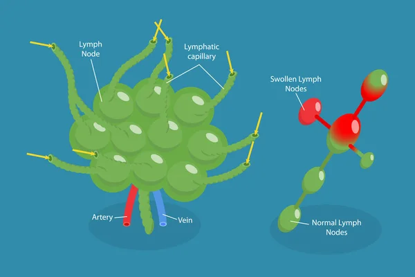
3D Isometric Flat Vector Conceptual Illustration Of Lymph Node, Labeled Diagram

Vector, 1.89MB, 6000 × 4000 eps
stock image Human body with cancer cells spreading and growing

Human Body With Cancer Cells Spreading And Growing

Image, 6.41MB, 2870 × 3023 jpg
stock image Cancer cells

Cancer Cells

Image, 4.89MB, 5000 × 3799 jpg
stock image A growing Cancer Tumor spreading on healthy Tissue

A Growing Cancer Tumor Spreading On Healthy Tissue

Image, 6.99MB, 5000 × 3333 jpg
stock image Pancreas Cancer Anatomy

Pancreas Cancer Anatomy

Image, 6.24MB, 5670 × 3467 jpg
stock image Micro-organisms under the microscope

Micro-organisms Under The Microscope

Image, 19.03MB, 5000 × 4000 jpg
stock image Human Cytotoxic T cell

Human Cytotoxic T Cell

Image, 3.37MB, 3840 × 2160 jpg
stock image Mesothelial malignant cells with multiple nuclei

Mesothelial Malignant Cells With Multiple Nuclei

Image, 2.18MB, 3658 × 3417 jpg
stock image Human Cytotoxic T cell

Human Cytotoxic T Cell

Image, 3.35MB, 3840 × 2160 jpg
stock image mesothelial cells in body fluid.

Mesothelial Cells In Body Fluid.

Image, 2.83MB, 3058 × 2957 jpg
stock image Mature lymphocyte on red blood cells medical background.

Mature Lymphocyte On Red Blood Cells Medical Background.

Image, 2.46MB, 3925 × 2507 jpg
stock image T-cells attacking a cancer cell. Isolated on black background. 3d render

T-cells Attacking A Cancer Cell. Isolated On Black Background. 3d Render

Image, 4.29MB, 4000 × 3000 jpg
stock vector Cancer and Neoplasm. comparison and difference between Malignant and Benign tumor. Benign tumor has a Capsule. Cells of Malignant tumor have Necrosis, Intravasation into the blood vessel, and Lymphatic Invasion.

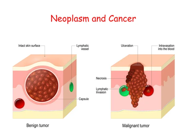
Cancer And Neoplasm. Comparison And Difference Between Malignant And Benign Tumor. Benign Tumor Has A Capsule. Cells Of Malignant Tumor Have Necrosis, Intravasation Into The Blood Vessel, And Lymphatic Invasion.

Vector, 12.6MB, 5000 × 3715 eps
stock image Extracapillary glomerulonephritis

Extracapillary Glomerulonephritis

Image, 1.6MB, 2450 × 2450 jpg
stock image Breast Tumor Symbol

Breast Tumor Symbol

Image, 10.81MB, 6600 × 6600 jpg
stock image Human Cytotoxic T cell

Human Cytotoxic T Cell

Image, 5.72MB, 3840 × 2160 jpg
stock image Abnormal malignant cells specimen pleural fluid.

Abnormal Malignant Cells Specimen Pleural Fluid.

Image, 6.15MB, 5186 × 3462 jpg
stock vector virus human papilloma

Virus Human Papilloma

Vector, 3.11MB, 6583 × 4458 eps
stock image Endometrial adenocarcinoma, light micrograph, photo under microscope

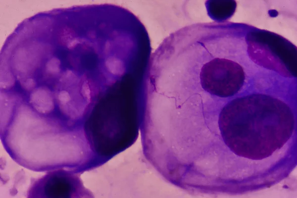
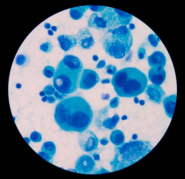
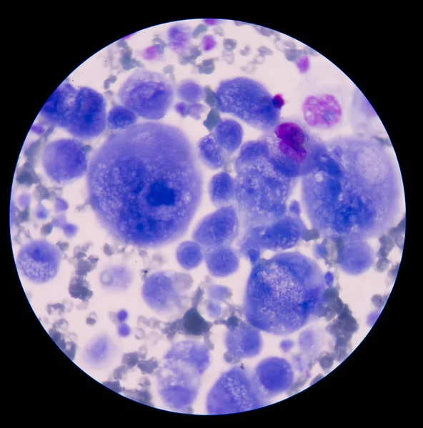
Endometrial Adenocarcinoma, Light Micrograph, Photo Under Microscope

Image, 14.51MB, 4386 × 2924 jpg
stock image Wilms tumor, or nephroblastoma, light micrograph, photo under microscope

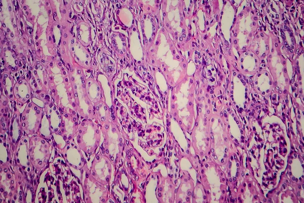
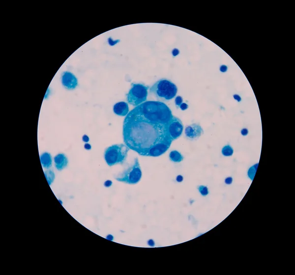
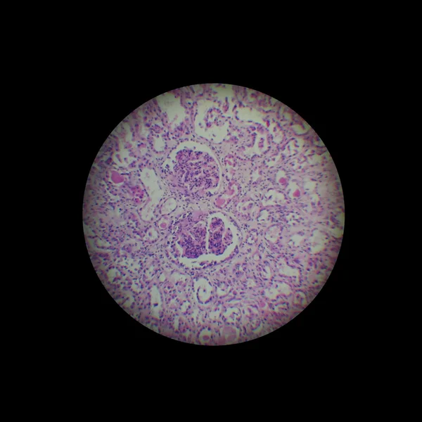
Wilms Tumor, Or Nephroblastoma, Light Micrograph, Photo Under Microscope

Image, 6.57MB, 3582 × 2388 jpg
stock image mesothelial cells in body fluid.

Mesothelial Cells In Body Fluid.

Image, 2.77MB, 2978 × 3017 jpg
stock vector Cancer prevention. Healthy cell and Cancer cell with DNA damage. carcinogens promote formation of malignant tumor by damage the genome. Probiotics, Prebiotics, and Synbiotics are Cancer prevention

Cancer Prevention. Healthy Cell And Cancer Cell With DNA Damage. Carcinogens Promote Formation Of Malignant Tumor By Damage The Genome. Probiotics, Prebiotics, And Synbiotics Are Cancer Prevention

Vector, 9.58MB, 4444 × 4444 eps
stock vector Cancer Devlopment Process infographic  illustration

Cancer Devlopment Process Infographic Illustration

Vector, 0.61MB, 6540 × 3611 eps
stock image PC-3 human prostate cancer cells stained with blue Coomassie, un

PC-3 Human Prostate Cancer Cells Stained With Blue Coomassie, Un

Image, 6.34MB, 4184 × 2789 jpg
stock image Moderate triple phosphate with bacteria in urine analysis medica

Moderate Triple Phosphate With Bacteria In Urine Analysis Medica

Image, 3.64MB, 2993 × 2974 jpg
stock image Malignant cells in pleural fluid.

Malignant Cells In Pleural Fluid.

Image, 6.04MB, 5215 × 3315 jpg
stock vector Structure of Melanocyte

Structure Of Melanocyte

Vector, 5.38MB, 5099 × 5099 eps
stock image malignant cells with multiple nuclei and prominent nucleoli.Meso

Malignant Cells With Multiple Nuclei And Prominent Nucleoli.Meso

Image, 2.77MB, 3000 × 3000 jpg
stock image breast cancer cell

Breast Cancer Cell

Image, 4.52MB, 3840 × 2160 jpg
stock image Uterine cancer, light micrograph

Uterine Cancer, Light Micrograph

Image, 15.04MB, 4180 × 2787 jpg
stock image mesothelial cells in body fluid.

Mesothelial Cells In Body Fluid.

Image, 1.73MB, 2958 × 2997 jpg
stock image Cancer cell in the moment that divides

Cancer Cell In The Moment That Divides

Image, 5.48MB, 5001 × 3865 jpg
stock vector Multiple myeloma. Close-up of Healthy bone marrow and Plasma cell myeloma. Red and White blood cells, normal and abnormal antibodies. cancer of plasma cells, that produces abnormal antibodies. Vector poster

Multiple Myeloma. Close-up Of Healthy Bone Marrow And Plasma Cell Myeloma. Red And White Blood Cells, Normal And Abnormal Antibodies. Cancer Of Plasma Cells, That Produces Abnormal Antibodies. Vector Poster

Vector, 7.1MB, 4444 × 4444 eps
Page 1 >> Next