
Stock image Microscopical

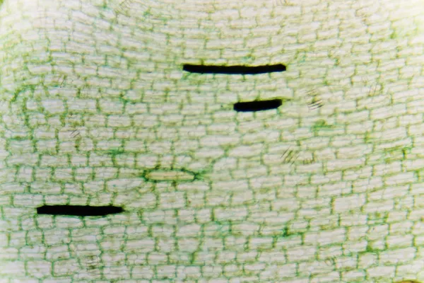
Microscopic Image Of Onion Root Tip Cells Undergoing Mitosis. Anaphases, Telophases And Metaphases. 1000x Magnified.
Image, 2.37MB, 2592 × 1944 jpg


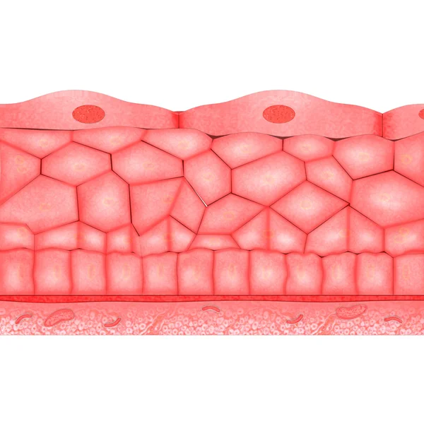
Red And White Blood Cell Character Design. Red Blood Cell Vector. Protective Mask.
Vector, 0.58MB, 9934 × 7016 eps


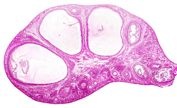
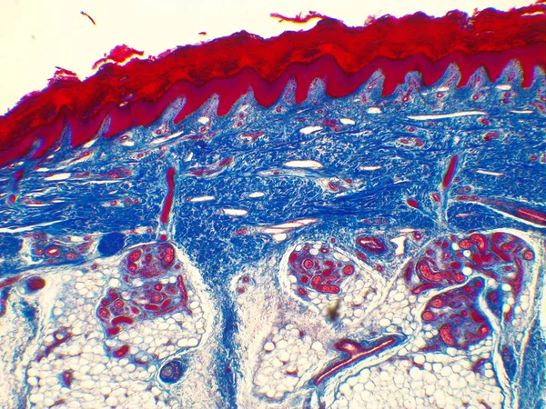
Micrograph Of Ovary Showing Primordial, Primary And Secondary Follicles Isolated On White Background.
Image, 13.12MB, 4624 × 3063 jpg

Microscopic Diagnosis Of Bacterial Vaginosis. Epithelial Cell, So-called Clue Cell Is Covered With Bacteria Gardnerella Vaginalis
Image, 2.53MB, 6000 × 4000 jpg
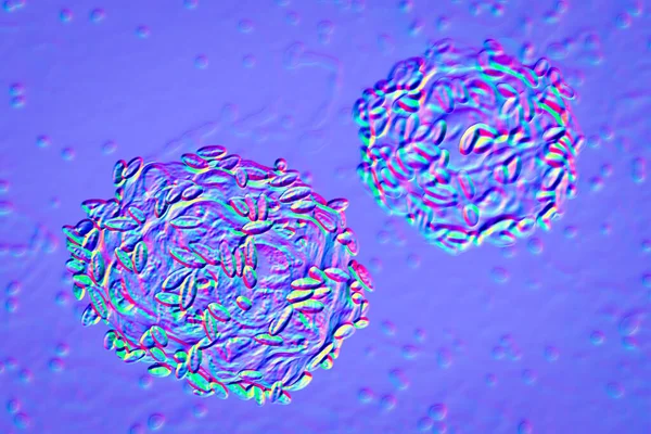
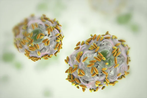
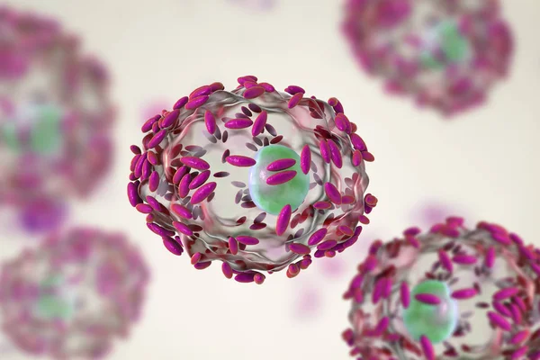
Bacterial Vaginosis. 3D Illustrations Showing Epithelial Cells Covered With Bacteria Gardnerella Vaginalis, So-called Clue Cells, Found In Vaginal Smear , 3D Illustration
Image, 7.41MB, 6000 × 4000 jpg


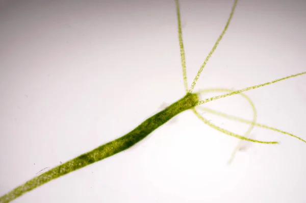
Hydra Is A Genus Of Small Fresh-water Animals Of The Phylum Cnidaria And Class Hydrozoa.
Image, 0MB, 5184 × 3456 jpg

Hydra Is A Genus Of Small Fresh-water Animals Of The Phylum Cnidaria And Class Hydrozoa.
Image, 0MB, 3456 × 5184 jpg


Erythrocytes, RBC. Vector Illustration Isolated On White Background.
Vector, 12.86MB, 5556 × 5556 eps

Chemistry Vector Chemical Science Or Pharmacy Research In School Laboratory For Technology Or Experiment In Laboratory Illustration Set Of Lab Scientific Equipment Microscope Isolated On Background
Vector, 0.84MB, 5000 × 5000 eps
Hydra Is A Genus Of Small, Fresh-water Animals Of The Phylum Cnidaria And Class Hydrozoa.
Image, 6.02MB, 4912 × 3264 jpg


Micrograph Of Ovary Showing Primordial, Primary And Secondary Follicles Isolated On White Background.
Image, 13.82MB, 4488 × 2972 jpg
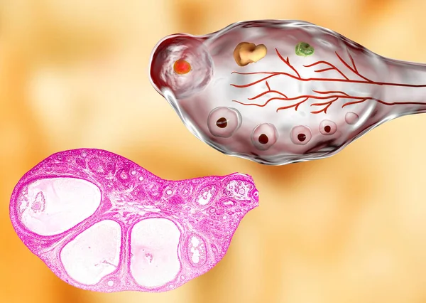
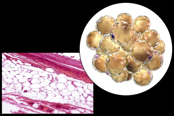
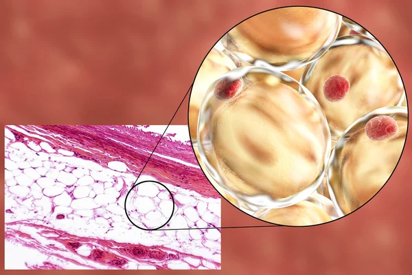
Transverse Section Of An Ovary Showing Primordial, Primary And Secondary Follicules. Light Microscopy, Hematoxylin And Eosin Stain, Magnification 200x And 3D Illustration
Image, 9.01MB, 5554 × 3948 jpg


Microscopic Image Of Onion Root Tip Cells Undergoing Mitosis. Anaphases And Metaphases. 1000x Magnified
Image, 2.36MB, 2592 × 1944 jpg

Hydra Is A Genus Of Small, Fresh-water Animals Of The Phylum Cnidaria And Class Hydrozoa.
Image, 3.9MB, 4912 × 3264 jpg

Hydra Is A Genus Of Small Fresh-water Animals Of The Phylum Cnidaria And Class Hydrozoa.
Image, 0MB, 5184 × 3456 jpg

Chemistry Vector Chemical Science Or Pharmacy Research In School Laboratory For Technology Or Experiment In Laboratory Illustration Set Of Lab Scientific Equipment Isolated On Background
Vector, 3.23MB, 5000 × 5000 eps

Microscopic Diagnosis Of Bacterial Vaginosis. Epithelial Cell, So-called Clue Cell Is Covered With Bacteria Gardnerella Vaginalis
Image, 4.15MB, 9000 × 6000 jpg
Microscopic Diagnosis Of Bacterial Vaginosis. Epithelial Cell, So-called Clue Cell Is Covered With Bacteria Gardnerella Vaginalis
Image, 3.01MB, 6000 × 4000 jpg
Microscopic Diagnosis Of Bacterial Vaginosis. Epithelial Cell, So-called Clue Cell Is Covered With Bacteria Gardnerella Vaginalis
Image, 4.07MB, 9000 × 6000 jpg


Page 1 >> Next