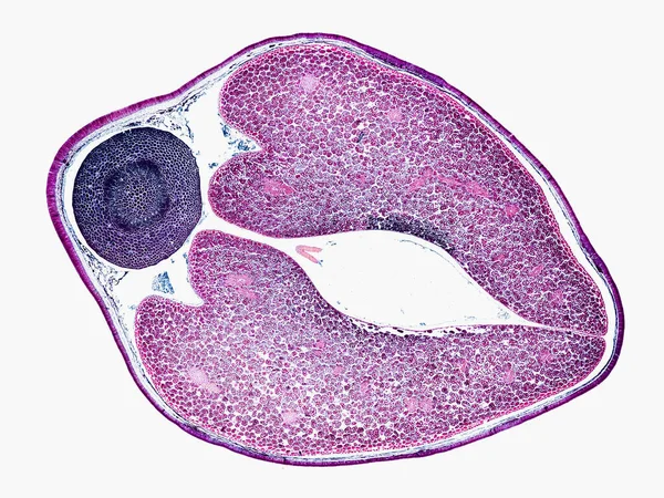
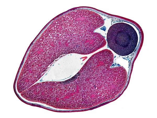
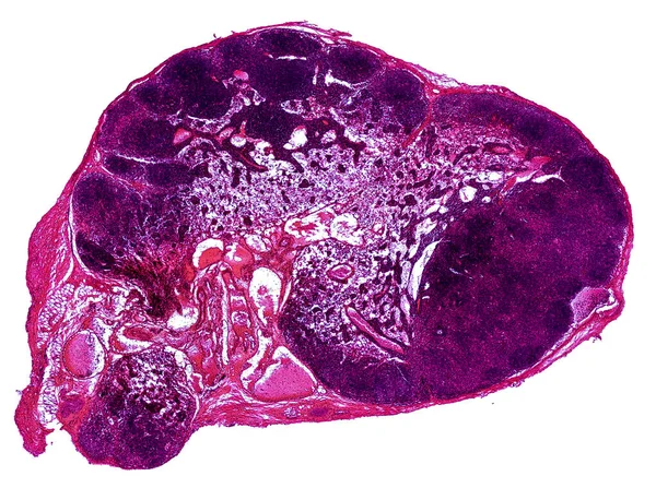
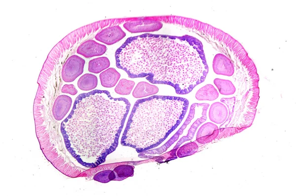
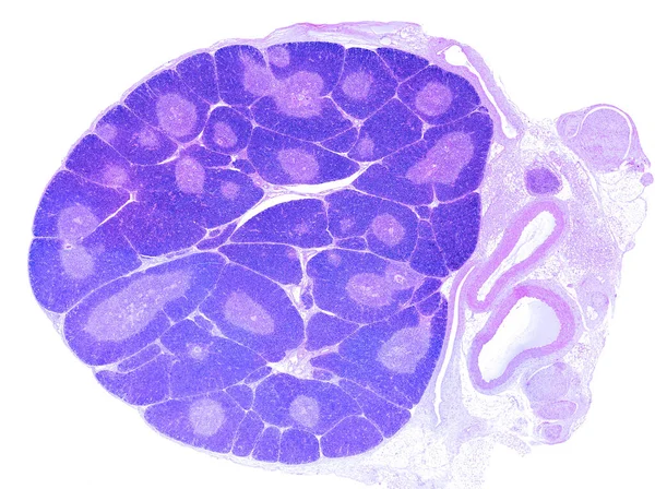
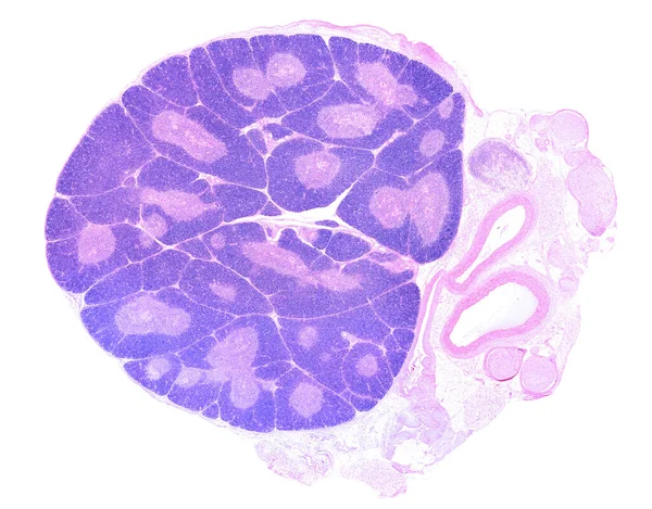
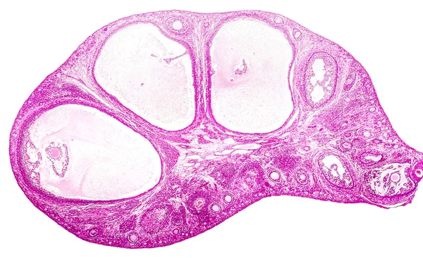
Stock image Light micrograph of ovary
Published: Sep.30, 2016 12:23:44
Author: katerynakon
Views: 47
Downloads: 1
File type: image / jpg
File size: 5.04 MB
Orginal size: 4120 x 2520 px
Available sizes:
Level: silver