photo free
Home Images Vectors Videos

Stock image Microscopie
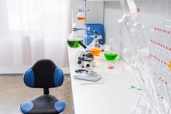
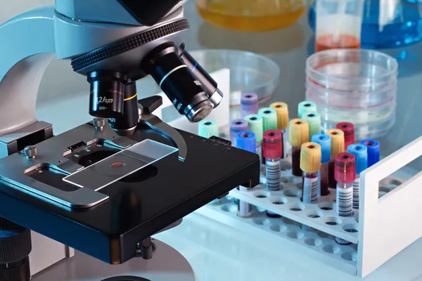
stock image Microscope and reagents in laboratory

Microscope And Reagents In Laboratory

Image, 12.11MB, 7360 × 4912 jpg
stock image close up of scientist looking to microscope in lab

Close Up Of Scientist Looking To Microscope In Lab

Image, 3.99MB, 3003 × 2002 jpg
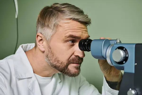
stock image A man meticulously examines someones vision.

A Man Meticulously Examines Someones Vision.

Image, 16.78MB, 8018 × 5345 jpg
stock vector Single flat microscope icon.

Single Flat Microscope Icon.

Vector, 0.21MB, 5000 × 5000 eps
stock image blood bank laboratory

Blood Bank Laboratory

Image, 9.95MB, 6144 × 4096 jpg
stock image Cancer cell in the moment that divides, 3d illustration

Cancer Cell In The Moment That Divides, 3d Illustration

Image, 6.82MB, 5001 × 3865 jpg
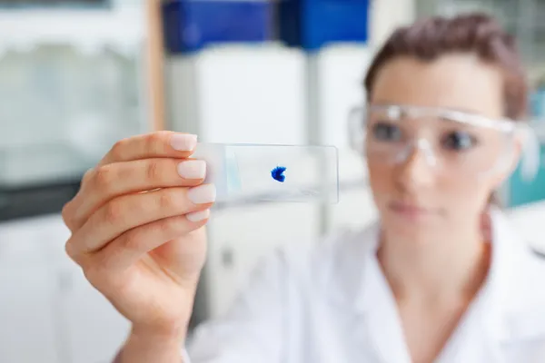
stock image close up of scientist hand with test sample in lab

Close Up Of Scientist Hand With Test Sample In Lab

Image, 5.71MB, 4245 × 2830 jpg
stock image Blood cells, medical or microbiological background with depth of field. 3d illustration

Blood Cells, Medical Or Microbiological Background With Depth Of Field. 3d Illustration

Image, 1.43MB, 5000 × 5000 jpg
stock image close up of scientist hand with test sample in lab

Close Up Of Scientist Hand With Test Sample In Lab

Image, 5.84MB, 4245 × 2830 jpg
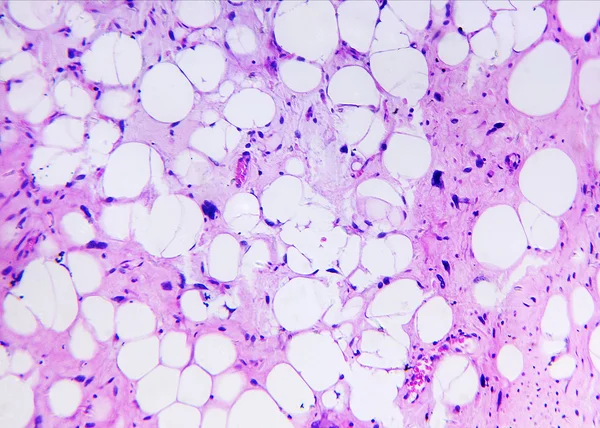
stock image Liposarcoma of a human

Liposarcoma Of A Human

Image, 4.01MB, 2370 × 1693 jpg
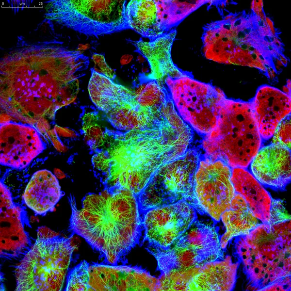
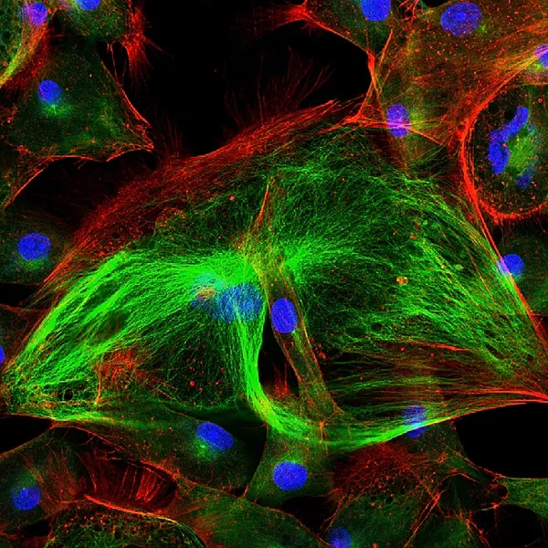
stock image Epithelial tumor cells labeled with fluorescent molecules

Epithelial Tumor Cells Labeled With Fluorescent Molecules

Image, 7.13MB, 3200 × 3200 jpg
stock image Background with fluorescent cancer cells

Background With Fluorescent Cancer Cells

Image, 1.02MB, 3200 × 3200 jpg
stock image microscope and glassware in laboratory

Microscope And Glassware In Laboratory

Image, 6.67MB, 5616 × 3744 jpg
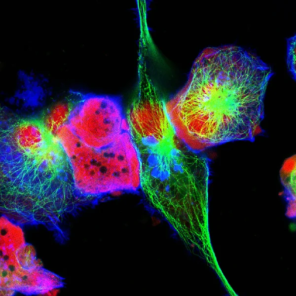
stock image Real fluorescence microscopic view of human neuroblastoma cells

Real Fluorescence Microscopic View Of Human Neuroblastoma Cells

Image, 3.76MB, 3000 × 3000 jpg
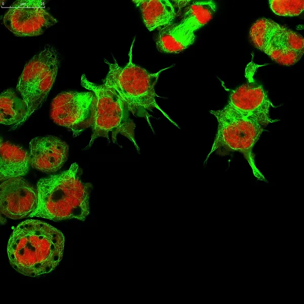
stock image Epithelial tumor cells labeled with fluorescent molecules

Epithelial Tumor Cells Labeled With Fluorescent Molecules

Image, 5.06MB, 2356 × 2356 jpg
stock image Microscope close-up on color background

Microscope Close-up On Color Background

Image, 7.17MB, 5760 × 3840 jpg
stock image Scientist looking at a microscope slide

Scientist Looking At A Microscope Slide

Image, 8.8MB, 5616 × 3744 jpg
stock image Scientist Using Urine Analyzer To Test Samples

Scientist Using Urine Analyzer To Test Samples

Image, 8.65MB, 5616 × 3744 jpg
stock image close up of scientist hand with test sample in lab

Close Up Of Scientist Hand With Test Sample In Lab

Image, 5.64MB, 4245 × 2830 jpg
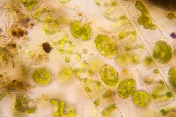
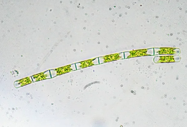
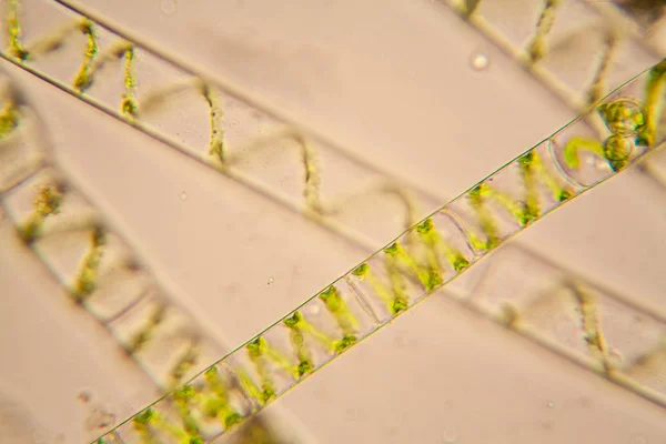
stock image Fresh pond water plankton and algae at the microscope. Spirogyra

Fresh Pond Water Plankton And Algae At The Microscope. Spirogyra

Image, 7.02MB, 4212 × 2808 jpg
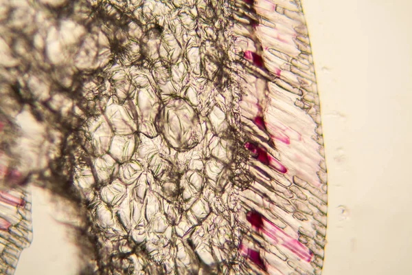
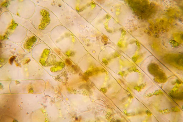
stock image Onion cells at the microscope

Onion Cells At The Microscope

Image, 7.17MB, 4212 × 2808 jpg
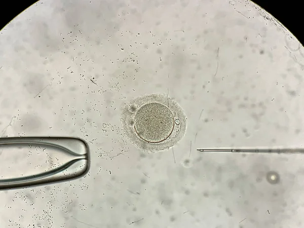
stock image View through microscope at in vitro fertilization process

View Through Microscope At In Vitro Fertilization Process

Image, 4.37MB, 4032 × 3024 jpg
stock image Laboratory Microscope. Scientific and healthcare research

Laboratory Microscope. Scientific And Healthcare Research

Image, 5.59MB, 4928 × 3264 jpg
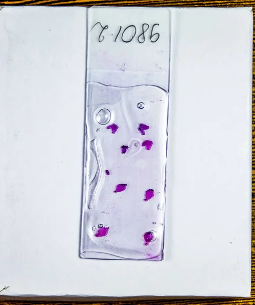
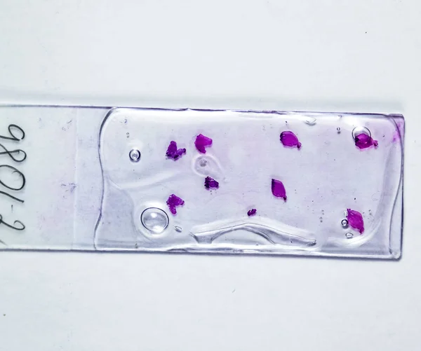
stock image Slices of the tumor under glass. Histological examination of tumor cells for the presence of cancer

Slices Of The Tumor Under Glass. Histological Examination Of Tumor Cells For The Presence Of Cancer

Image, 2.4MB, 2340 × 2800 jpg
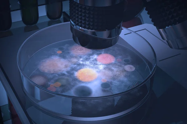
stock image Bacteria and fungus in a petri dish.

Bacteria And Fungus In A Petri Dish.

Image, 7.1MB, 4680 × 3120 jpg
stock image Algae under a microscope. The microscopic world.

Algae Under A Microscope. The Microscopic World.

Image, 6.03MB, 3835 × 2608 jpg
stock image Fresh pond water plankton and algae at the microscope. Spirogyra

Fresh Pond Water Plankton And Algae At The Microscope. Spirogyra

Image, 6.96MB, 4212 × 2808 jpg
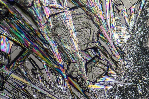
stock image soda lye microcrystals

Soda Lye Microcrystals

Image, 7.13MB, 3060 × 2040 jpg
stock image Fresh pond water plankton and algae at the microscope. Spirogyra

Fresh Pond Water Plankton And Algae At The Microscope. Spirogyra

Image, 6.28MB, 4212 × 2808 jpg
stock image close up of scientist hand with test sample in lab

Close Up Of Scientist Hand With Test Sample In Lab

Image, 5.92MB, 4245 × 2830 jpg
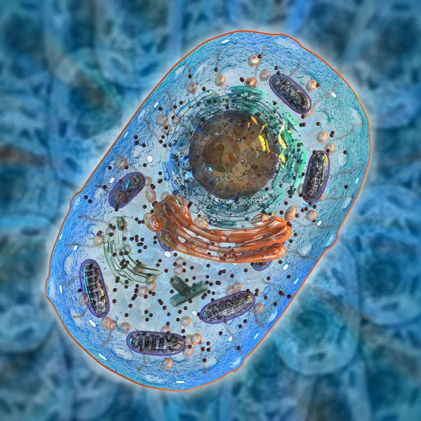
stock image Animal cell. Internal structure.

Animal Cell. Internal Structure.

Image, 14.09MB, 5000 × 5000 jpg
stock image Green and red chemical compounds microscope and test tubes in bi

Green And Red Chemical Compounds Microscope And Test Tubes In Bi

Image, 13.3MB, 4752 × 3168 jpg
stock image Finding parasites in feces in laboratory.

Finding Parasites In Feces In Laboratory.

Image, 13.53MB, 6720 × 4480 jpg
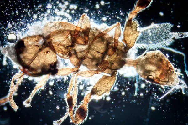
stock image animal insect

Animal Insect

Image, 9.15MB, 4752 × 3168 jpg
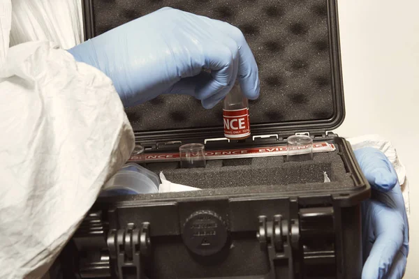
stock image Proccess of collecting possible gunshot residue from suspected man indoor - packing of evidence

Proccess Of Collecting Possible Gunshot Residue From Suspected Man Indoor - Packing Of Evidence

Image, 4.84MB, 5286 × 3524 jpg
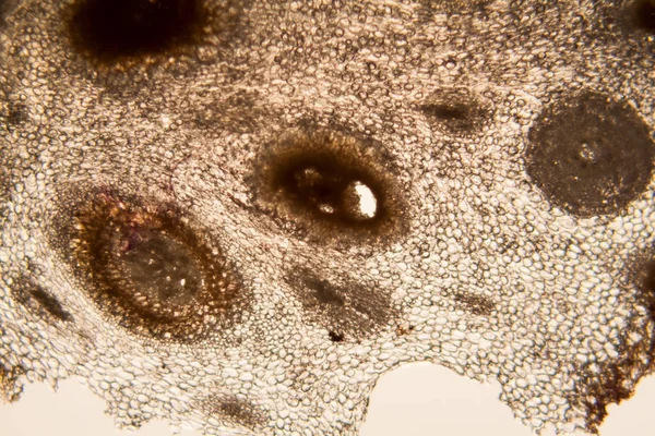
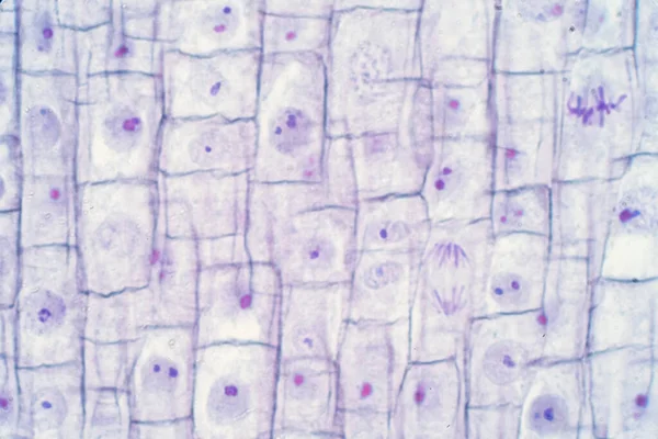
stock image onion root cells at the microscope

Onion Root Cells At The Microscope

Image, 13.45MB, 5616 × 3744 jpg
stock image science microscopy animal insect

Science Microscopy Animal Insect

Image, 7.25MB, 3264 × 3264 jpg
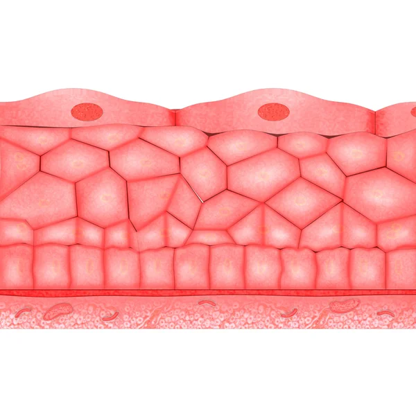
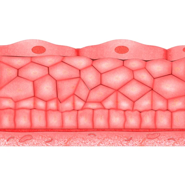
stock image Compound epithelium

Compound Epithelium

Image, 3.66MB, 4096 × 4096 jpg
stock image Cells in reproductive female cytology and histology concept.

Cells In Reproductive Female Cytology And Histology Concept.

Image, 1.64MB, 2884 × 2595 jpg
stock image Slices of the tumor under glass. Histological examination of tumor cells for the presence of cancer

Slices Of The Tumor Under Glass. Histological Examination Of Tumor Cells For The Presence Of Cancer

Image, 2.35MB, 2800 × 2340 jpg
stock image Scientific Microscopic photography. Mold.

Scientific Microscopic Photography. Mold.

Image, 1.55MB, 3476 × 2318 jpg
stock image Human epithelium anatomy

Human Epithelium Anatomy

Image, 3.66MB, 4096 × 4096 jpg
stock image Root tip of Onion and Mitosis cell in the Root tip for education

Root Tip Of Onion And Mitosis Cell In The Root Tip For Education

Image, 18.88MB, 6000 × 4000 jpg
stock image Slide show Parasite Strongyloides stercoralis in sputum smear.

Slide Show Parasite Strongyloides Stercoralis In Sputum Smear.

Image, 2.81MB, 2989 × 2837 jpg
stock image soda lye microcrystals

Soda Lye Microcrystals

Image, 5.91MB, 3060 × 2040 jpg
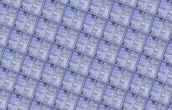
stock image Detail of a silicon wafer

Detail Of A Silicon Wafer

Image, 20.35MB, 5000 × 3214 jpg
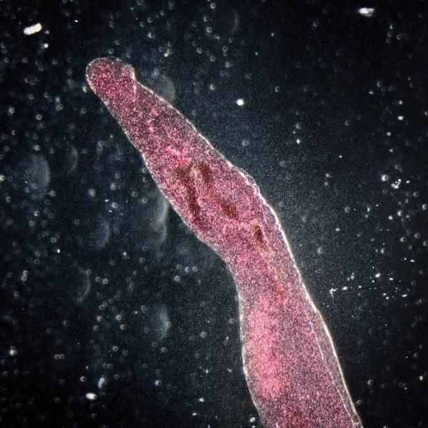
stock image animal parasiteras schistosome blood flukes

Animal Parasiteras Schistosome Blood Flukes

Image, 6.39MB, 3264 × 3264 jpg
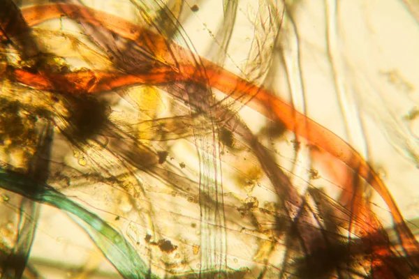
stock image Microscopic view of dust

Microscopic View Of Dust

Image, 11.09MB, 5616 × 3744 jpg
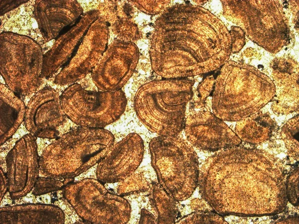
stock image  Micro-photo of geological thin section under cross-polarized light.

Micro-photo Of Geological Thin Section Under Cross-polarized Light.

Image, 9.85MB, 4724 × 3543 jpg
stock image Abstract biological pattern

Abstract Biological Pattern

Image, 3.58MB, 2560 × 1920 jpg
Page 1 >> Next