
Stock image Microscopie page 2


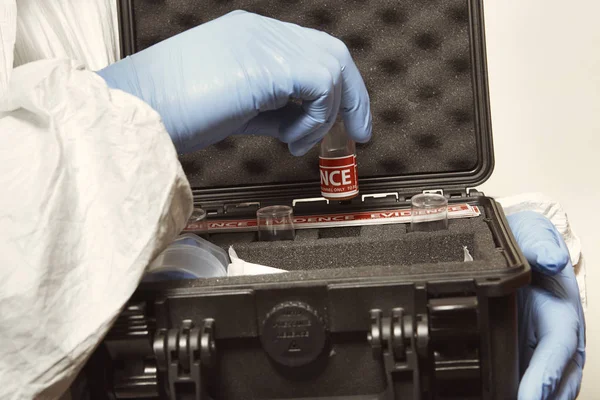
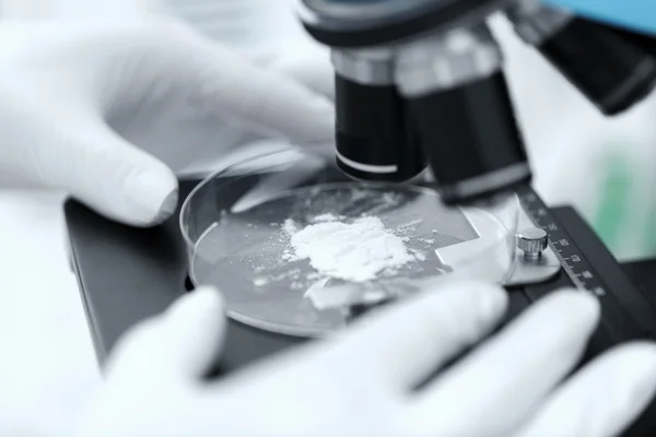
Proccess Of Collecting Possible Gunshot Residue From Suspected Man Indoor - Packing Of Evidence
Image, 4.84MB, 5286 × 3524 jpg
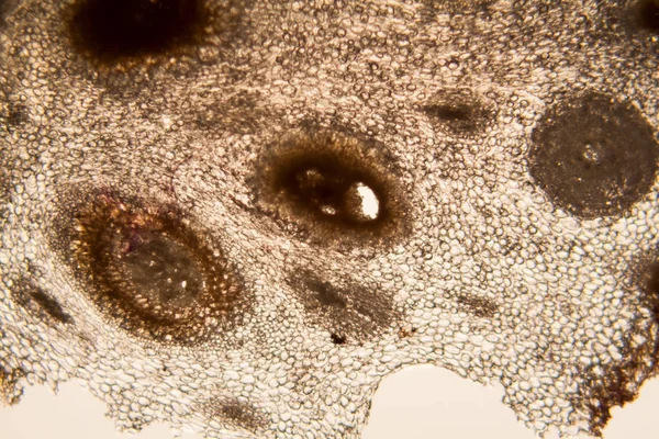


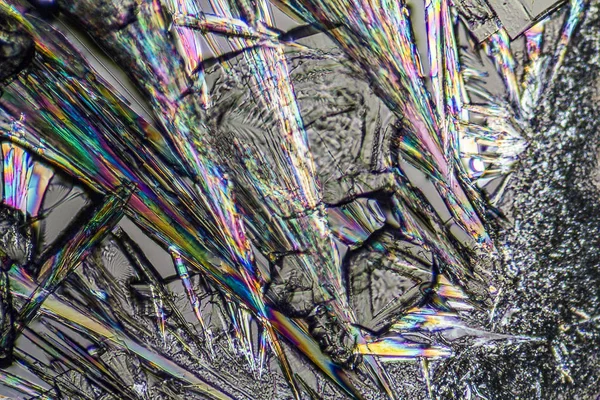
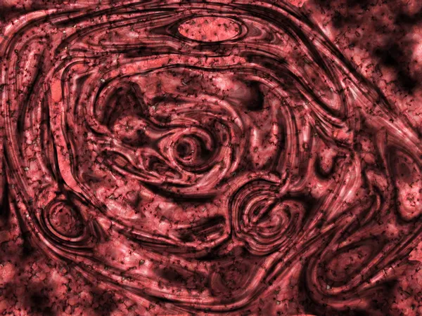
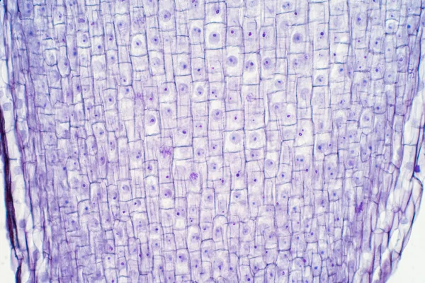
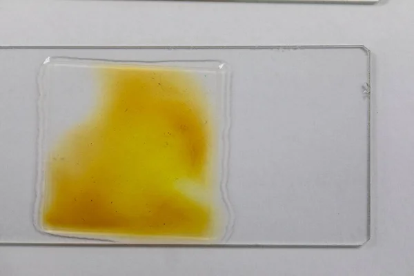
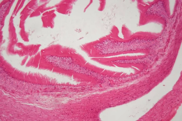
Slices Of The Tumor Under Glass. Histological Examination Of Tumor Cells For The Presence Of Cancer
Image, 2.35MB, 2800 × 2340 jpg


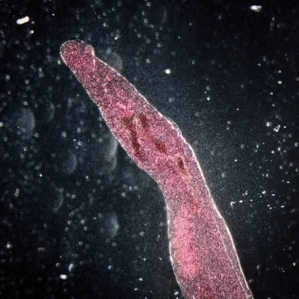

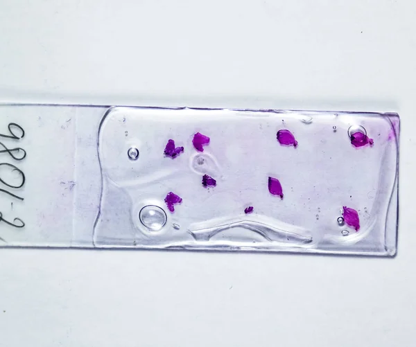
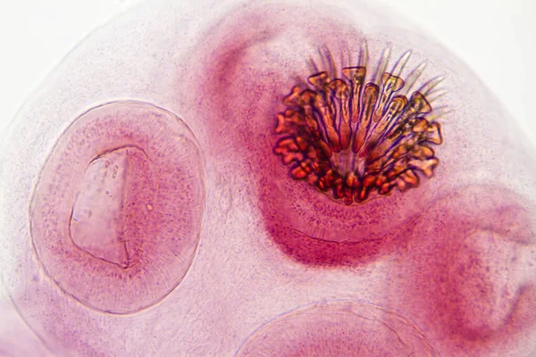
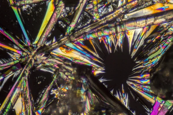
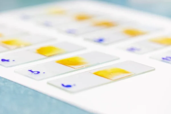
Slices Of The Tumor Under Glass. Histological Examination Of Tumor Cells For The Presence Of Cancer
Image, 2.75MB, 4000 × 3094 jpg


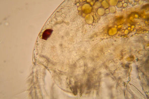
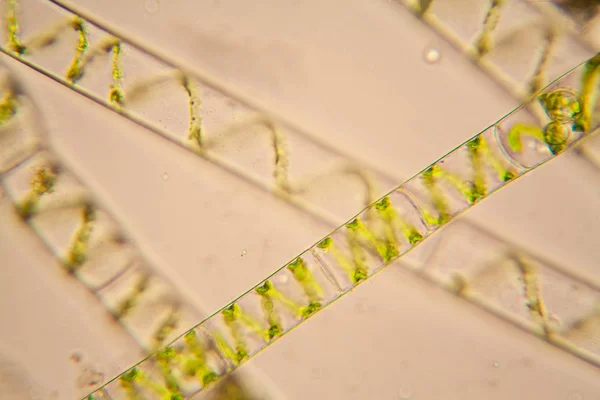
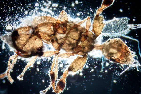
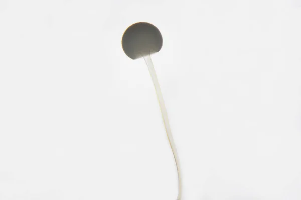
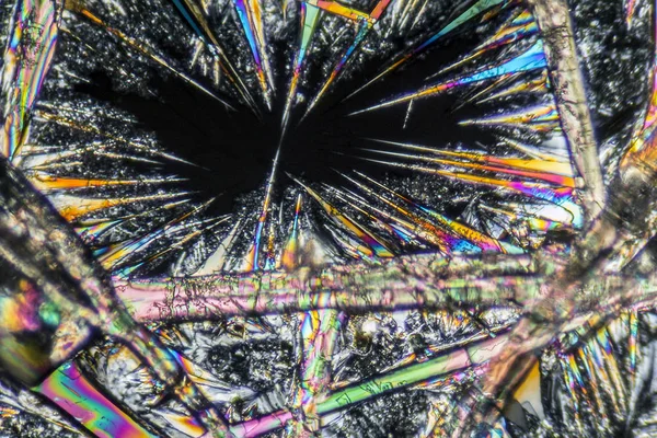
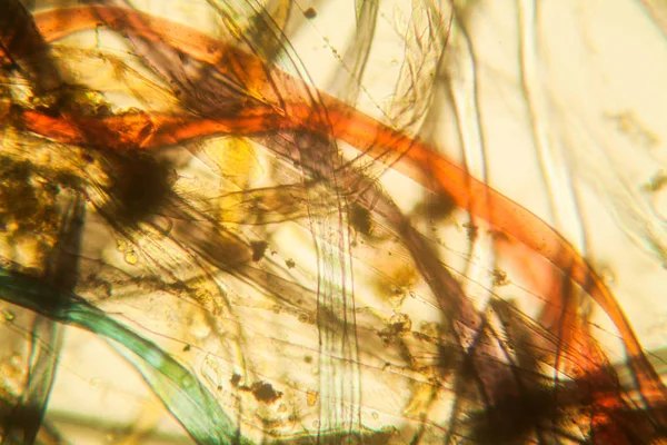
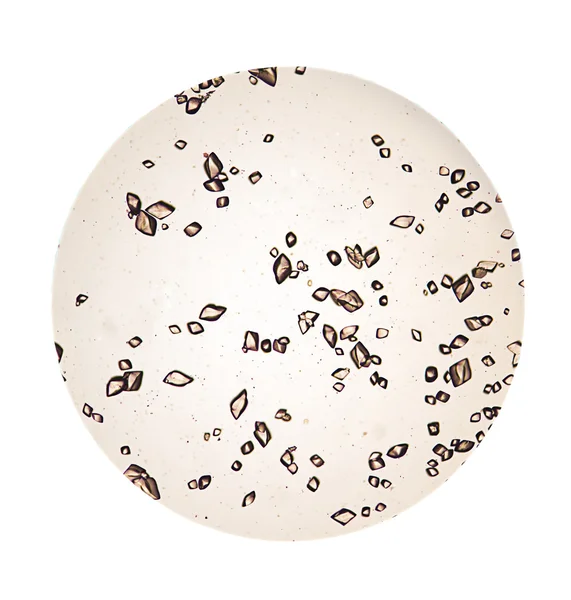
Fresh Pond Water Plankton And Algae At The Microscope. Copepod Details
Image, 6.57MB, 4212 × 2808 jpg
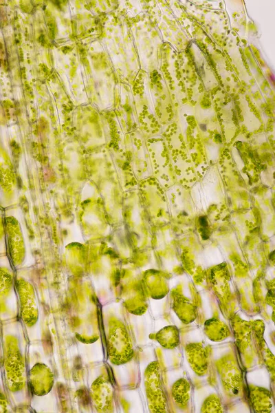
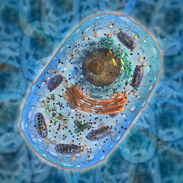
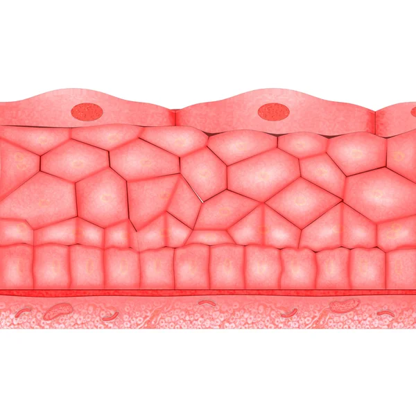
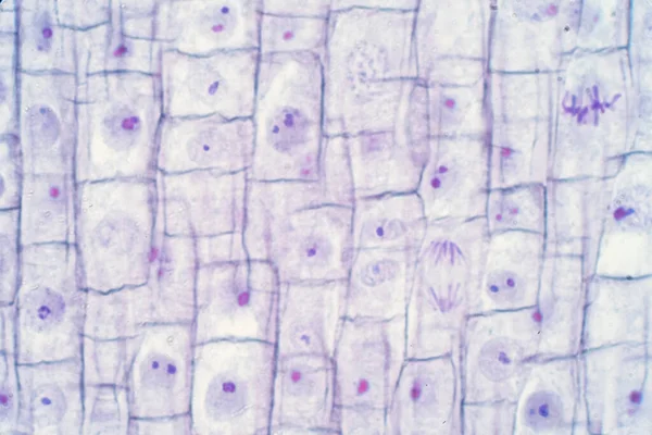
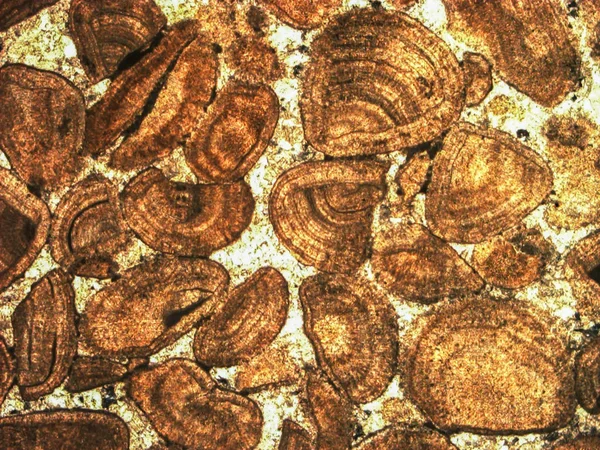
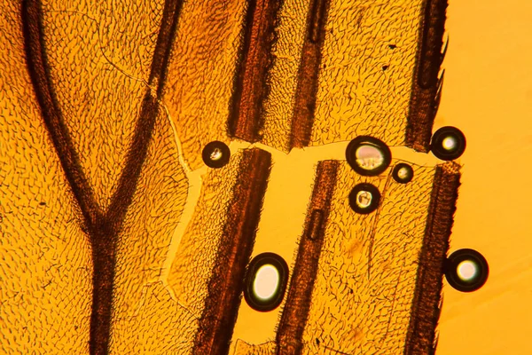
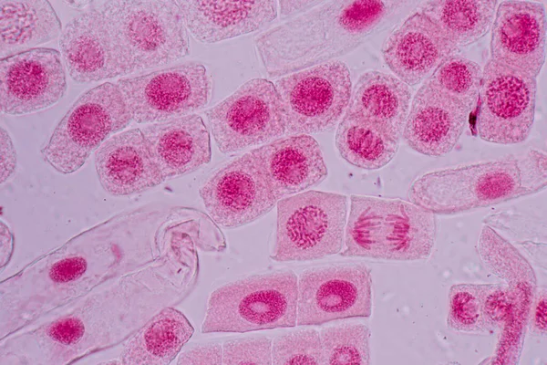
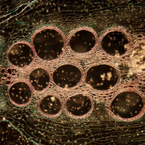
Cell Structure Hydrilla, View Of The Leaf Surface Showing Plant Cells Under The Microscope For Classroom Education.
Image, 6.97MB, 2560 × 3840 jpg


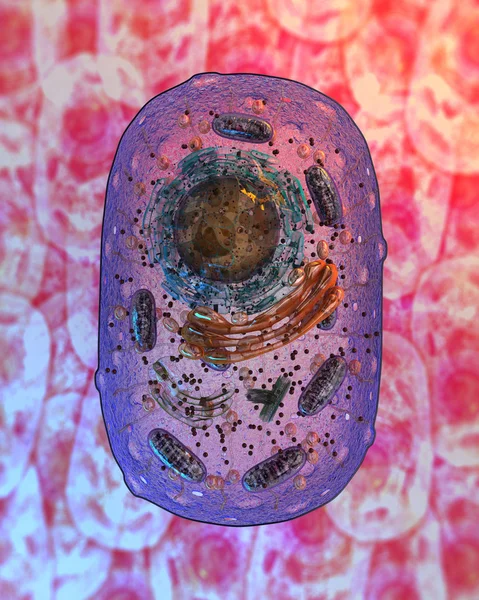
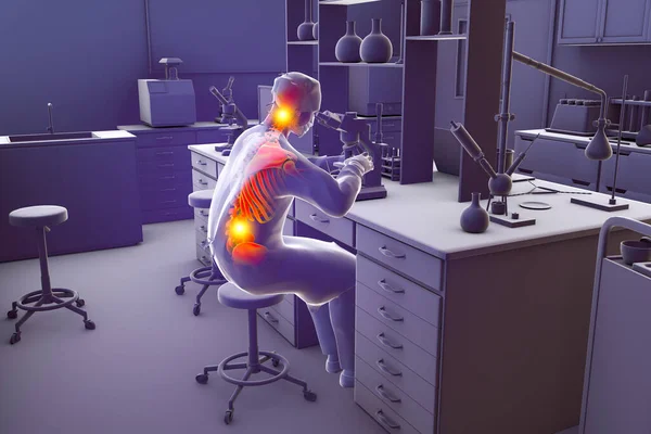
Work-related Musculoskeletal Disorders In Laboratory Workers, Conceptual 3D Illustration Showing A Lab Specialist With Highlighted Skeleton Working With Microscope Having Neck And Back Pain
Image, 8.55MB, 6000 × 4000 jpg


Previous << Page 2 >> Next