
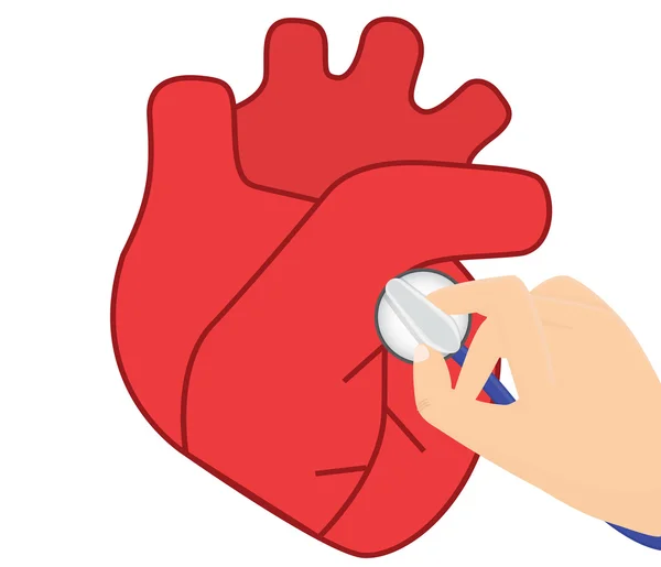
Stock image Mitral Valve Stenosis

Mitral Stenosis. A Narrowing Of The Mitral Orifice, Obstructing The Flow Of Blood From The Left Atrium To The Left Ventricle. Simple Black And White Vector Illustration.
Vector, 0.4MB, 5650 × 2985 eps
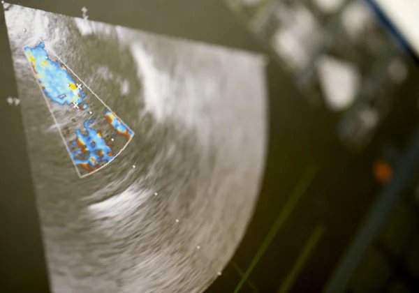
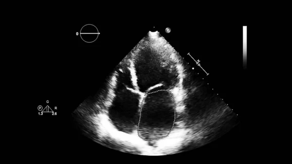
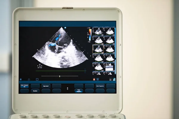
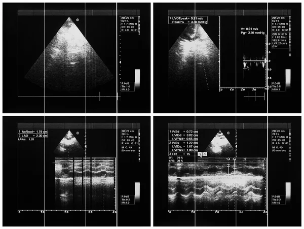
Image Of The Heart In Gray-scale Mode During Transesophageal Ultrasound.
Image, 1.19MB, 4000 × 2250 jpg


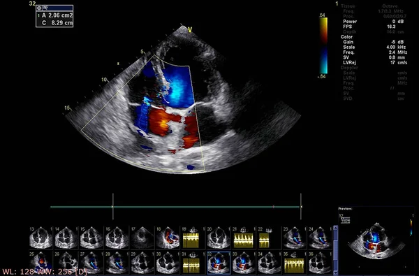

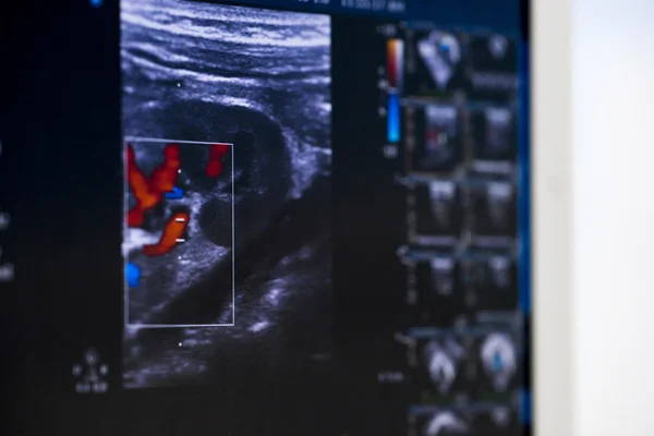
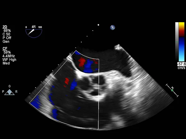
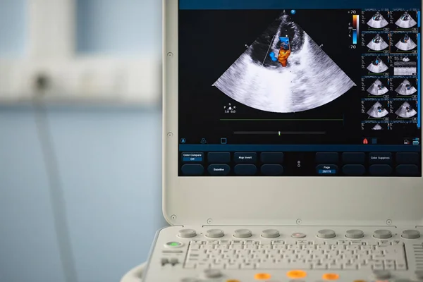
On The Screen Of A Modern Ultrasound Scanner, An Image Of The Heart Cavities With Red And Blue Streams Of Blood Regurgitation Depicted By The Doppler Method.
Image, 10.59MB, 5500 × 3670 jpg
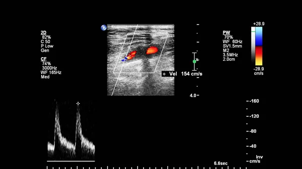
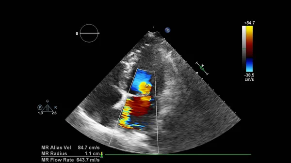

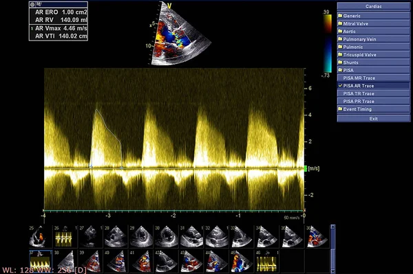

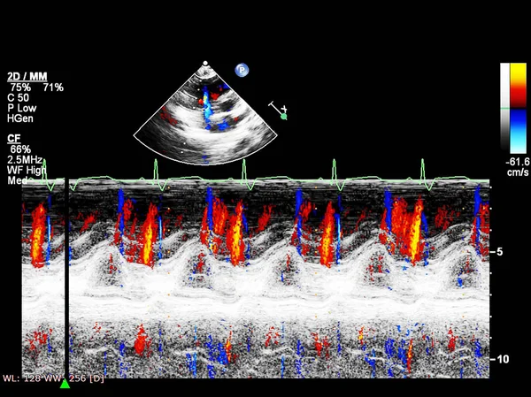


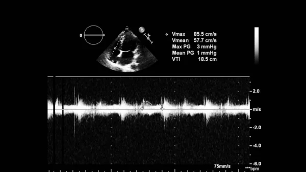
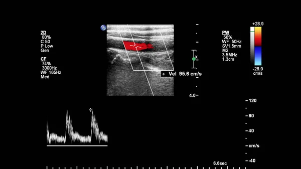
Image Of The Heart During Transesophageal Ultrasound With Doppler Mode.
Image, 0.61MB, 4000 × 2250 jpg
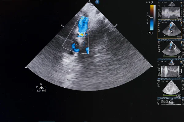
On The Ultrasound Screen, The Image Of The Heart In The Four-chamber Position And The Doppler Method Shows Tricuspid Regurgitation.
Image, 12.68MB, 5500 × 3671 jpg


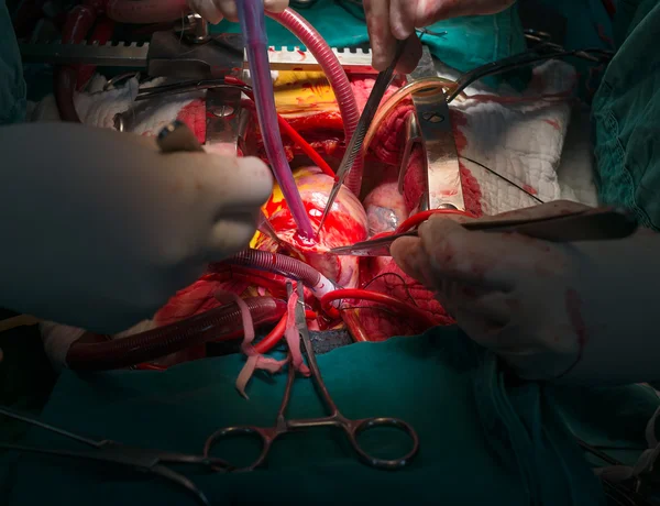
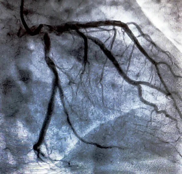
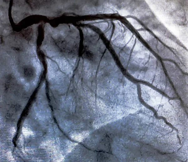
Coronary Artery Angiogram Of Left Coronary Artery During Cardiac Catheterization . Catheterization. Cardiac Ventriculography Is A Medical Imaging Test Used To Determine A Patient Cardiac Function
Image, 1.49MB, 3016 × 2890 jpg
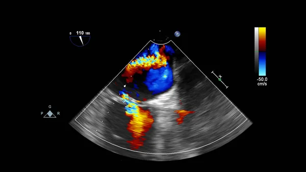
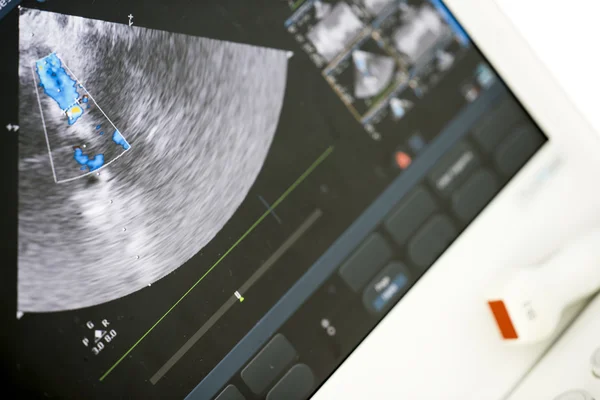
Image Of The Heart During Transesophageal Ultrasound With Doppler Mode.
Image, 1.05MB, 3500 × 1969 jpg

Catheterization. Cardiac Ventriculography Is A Medical Imaging Test Used To Determine A Patient Cardiac Function In The Right Or Left Ventricle
Image, 1.16MB, 3874 × 2071 jpg

Catheterization. Cardiac Ventriculography Is A Medical Imaging Test Used To Determine A Patient Cardiac Function In The Right Or Left Ventricle
Image, 2.42MB, 3007 × 2930 jpg
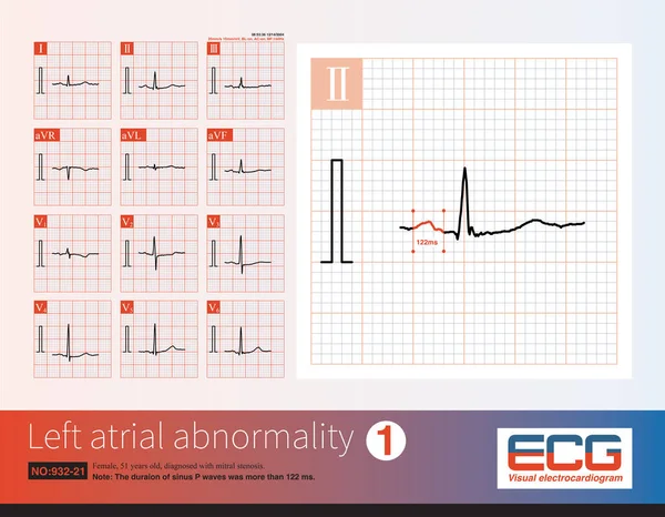
Female, 51 Years Old, Diagnosed With Mitral Stenosis. When This ECG Was Taken, The Patient Still Maintained Sinus Rhythm.Note That The P Wave Duration Was Widened.
Image, 14.21MB, 10000 × 7772 jpg

Coronary Artery Angiogram Of Left Coronary Artery During Cardiac Catheterization . Catheterization. Cardiac Ventriculography Is A Medical Imaging Test Used To Determine A Patient Cardiac Function
Image, 1.12MB, 3001 × 2590 jpg


On The Screen Of A Modern Ultrasound Scanner, An Image Of The Heart Cavities With Red And Blue Streams Of Blood Regurgitation Depicted By The Doppler Method.
Image, 12.45MB, 6000 × 4005 jpg

Catheterization. Cardiac Ventriculography Is A Medical Imaging Test Used To Determine A Patient Cardiac Function In The Right Or Left Ventricle
Image, 1.4MB, 3028 × 2890 jpg

Coronary Artery Angiogram Of Left Coronary Artery During Cardiac Catheterization . Catheterization. Cardiac Ventriculography Is A Medical Imaging Test Used To Determine A Patient Cardiac Function
Image, 4.97MB, 2582 × 2500 jpg

Coronary Artery Angiogram Of Left Coronary Artery During Cardiac Catheterization . Catheterization. Cardiac Ventriculography Is A Medical Imaging Test Used To Determine A Patient Cardiac Function
Image, 1.34MB, 3071 × 2650 jpg
On The Screen Of The Ultrasound Scanner, The Heart Image In The Four-chamber Position And Marked By The Blue And Red Doppler Method Are Valve Regurgitation.
Image, 12.07MB, 5500 × 3671 jpg

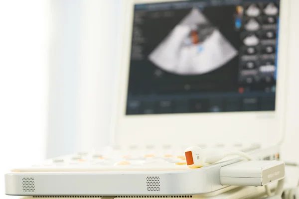
Image Of The Heart In Gray-scale Mode During Transesophageal Ultrasound.
Image, 0.63MB, 4000 × 2250 jpg
Page 1 >> Next