
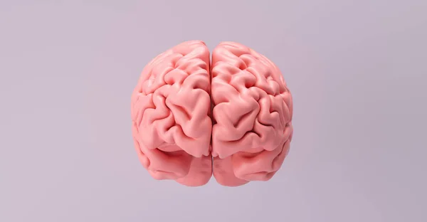
Stock image Motor Cortex
Human Brain With Highlighted Precentral And Postcentral Gyri, 3D Illustration. The Sites Of Primary Motor (precentral Gyrus) And Somatosensory (postcentral Gyrus) Cortex
Image, 3.12MB, 6000 × 4000 jpg
Areas Of The Brain: Prefrontal Cortex, Premotor Cortex And Oculomotor Cortex. .
Image, 0.93MB, 3630 × 3042 jpg


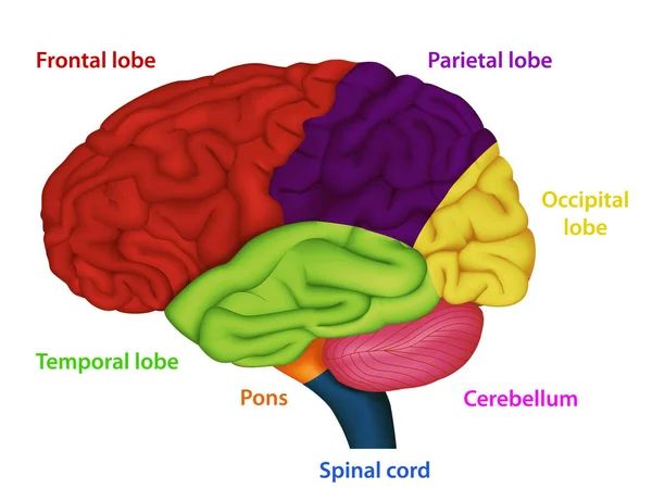
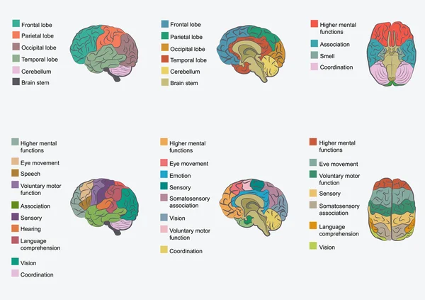
Areas Of The Human Brain ,medical Vector Illustration On White Background
Vector, 5MB, 6500 × 5000 eps
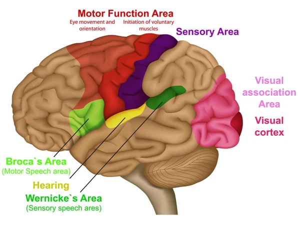
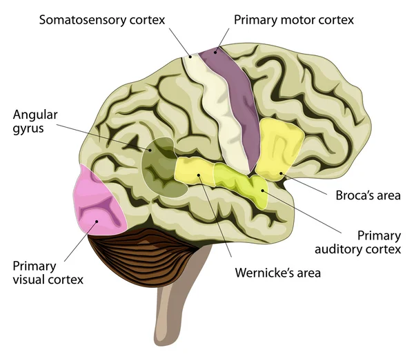
Functional Brain Areas Medical Vector Illustration On White Background
Vector, 4.29MB, 6500 × 5000 eps
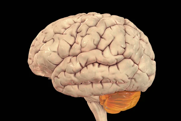
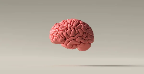
Human Brain With Highlighted Cerebellum, 3D Illustration. It Plays An Important Role In Motor Control And Is Involved In Some Cognitive Functions, Attention, Language, Emotional Control
Image, 4.85MB, 6166 × 4111 jpg

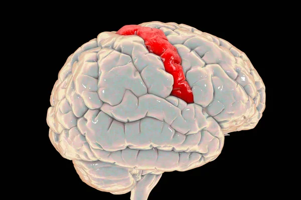
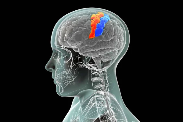
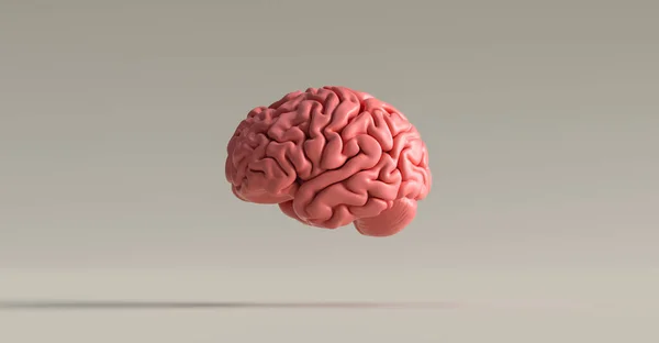
Human Brain With Highlighted Precentral Gyrus, Side View, 3D Illustration. It Is Located In The Posterior Frontal Lobe And Is The Site Of The Primary Motor Cortex, The Brodmann Area 4.
Image, 5.54MB, 6000 × 4000 jpg
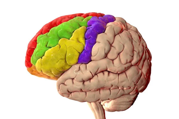
Human Brain With Highlighted Frontal Gyri, Superior (red), Middle (green), Inferior (yellow), Precentral (blue), 3D Illustration
Image, 5.09MB, 6000 × 4000 jpg
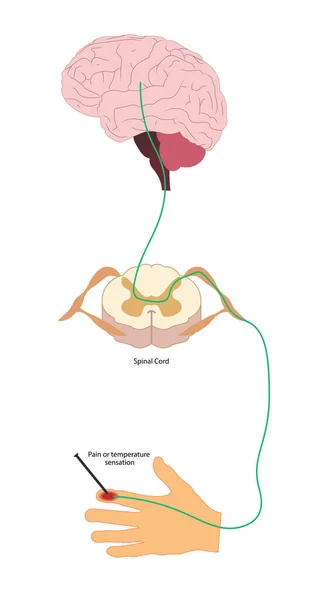
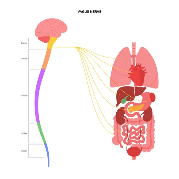
Pain Pathway. Nociception. Ascending Pathway That Connect The Periphery With The Brain During Pain And Temperature Sensation. Hand, Spinal Cord And Brain.
Image, 3.76MB, 7230 × 14001 jpg
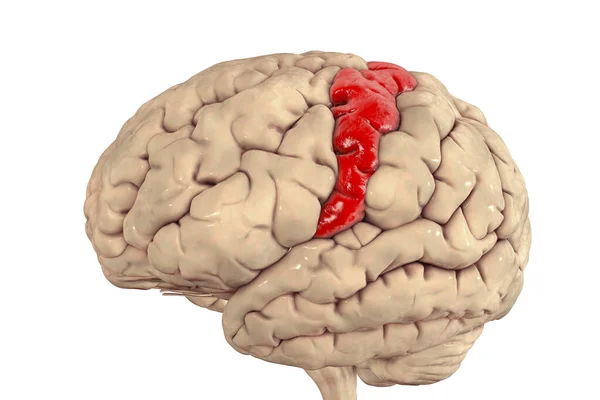
Human Brain With Highlighted Precentral Gyrus, 3D Illustration. It Is Located In The Posterior Frontal Lobe And Is The Site Of The Primary Motor Cortex, The Brodmann Area 4.
Image, 4.54MB, 6100 × 4066 jpg
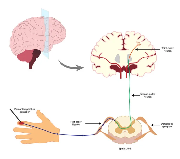
Pain Pathway. Nociception. Ascending Pathway That Connect The Periphery With The Brain During Pain And Temperature Sensation. Hand, Spinal Cord And Brain.
Image, 2.2MB, 6192 × 5490 jpg

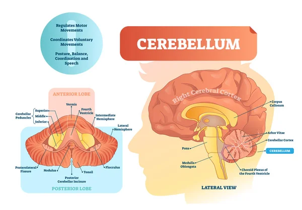
Cerebellum Vector Illustration. Medical Labeled Diagram With Internal View.
Vector, 7.74MB, 5000 × 3491 eps
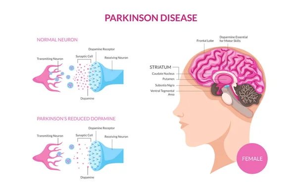
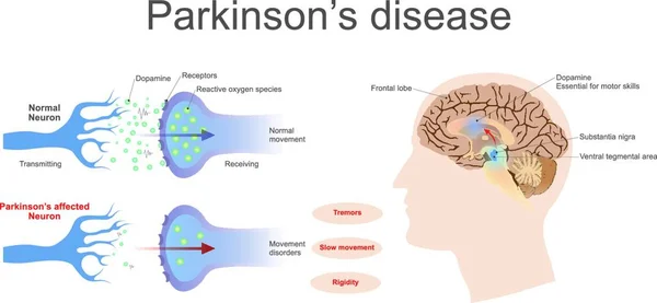
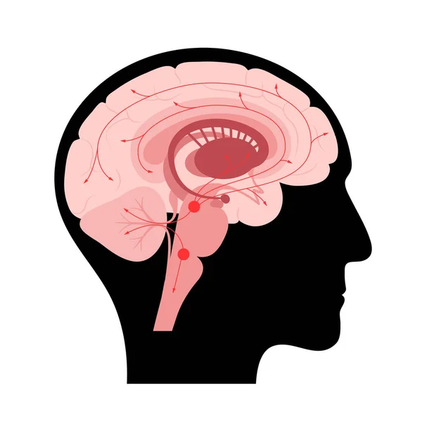
Parkinson Disease On Female Patient Detail Medical Illustration, Suitable For Medical Poster, Awareness Campaign, Editorial, Print, And Other Health Related Occasion
Vector, 6.33MB, 9567 × 6250 eps


Human Brain With Highlighted Cerebellum And Close-up View Of Purkinje Neurons, One Of The Commonest Types Of Cells In Cerebellar Cortex, 3D Illustration
Image, 10.04MB, 8029 × 4517 jpg

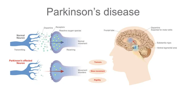
Parkinson Disease On Male Patient Detail Medical Illustration, Suitable For Medical Poster, Awareness Campaign, Editorial, Print, And Other Health Related Occasion
Vector, 6.32MB, 9567 × 6250 eps

Coronal Section Of A Healthy Brain Showing Normal Anatomy Of Basal Baglia And Ventricles, 3D Illustration
Image, 4.88MB, 6000 × 4000 jpg

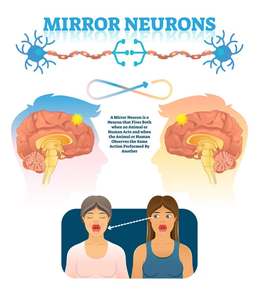
Mirror Neurons Vector Illustration. Medical Brain Action Explanation Scheme
Vector, 9.28MB, 4000 × 4519 eps
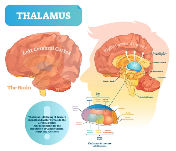
Thalamus Vector Illustration. Labeled Medical Diagram With Brain Structure.
Vector, 13.72MB, 4500 × 3873 eps
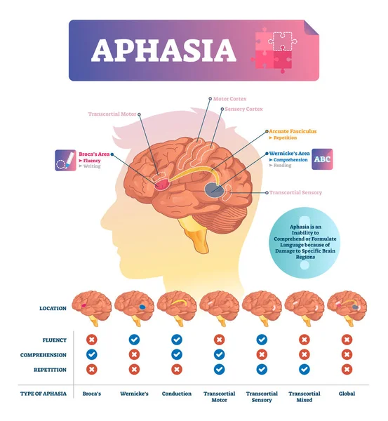
Aphasia Vector Illustration. Labeled Educational Scheme With Brain Disorder
Vector, 8.09MB, 4000 × 4335 eps

Human Brain With Highlighted Cerebellum And Close-up View Of Purkinje Neurons, One Of The Commonest Types Of Cells In Cerebellar Cortex, 3D Illustration
Image, 11.25MB, 8029 × 4517 jpg

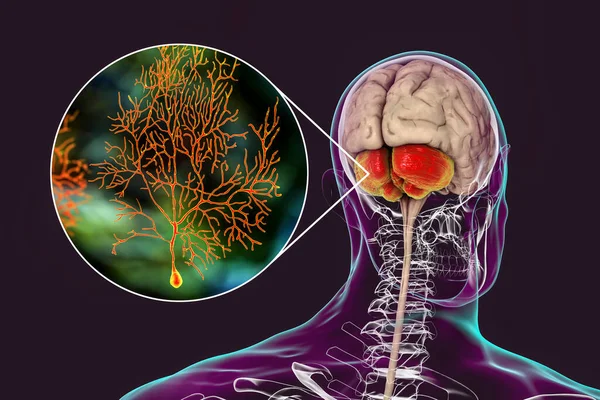
Human Brain With Highlighted Cerebellum And Close-up View Of Purkinje Neurons, One Of The Commonest Types Of Cells In Cerebellar Cortex, 3D Illustration
Image, 7.54MB, 6000 × 4000 jpg
Page 1 >> Next