
Stock image Motor Neuron Disease

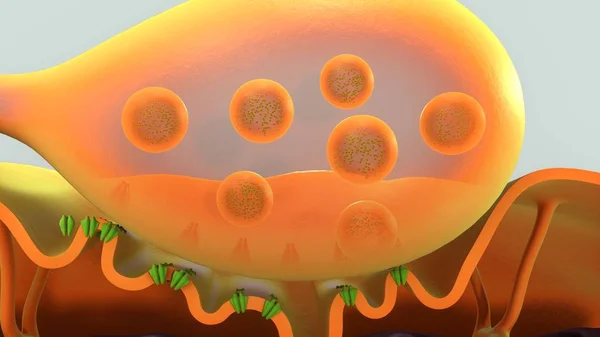
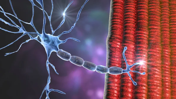
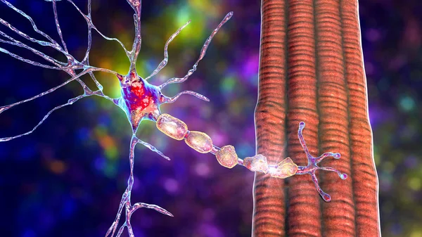
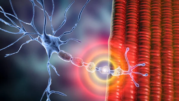
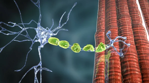
Motor Neuron Connecting To Muscle Fiber, 3D Illustration. A Neuromuscular Junction Allows The Motor Neuron To Transmit A Signal To The Muscle Causing Contraction. It Is Affected By Toxins And Diseases
Image, 11.3MB, 7200 × 4050 jpg

Neurological Physical Examination Of The Hands Reflexes. Doctor Neurologist Checks The Status Of The Patient's Reflexes In Office In Hospital. Selective Focus, Space For Text.
Image, 8.47MB, 5767 × 3850 jpg
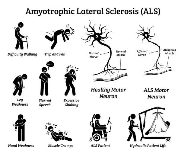
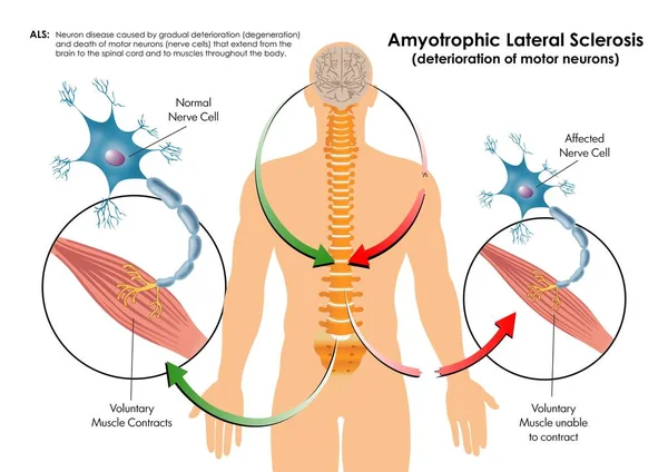
Amyotrophic Lateral Sclerosis ALS Disease Signs And Symptoms. Illustrations Depict Nervous System Or Neurological Disease In ALS Patient.
Vector, 4.71MB, 7500 × 6500 eps
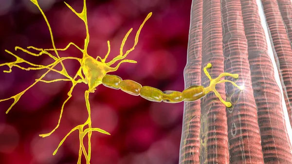
Motor Neuron Connecting To Muscle Fiber, 3D Illustration. A Neuromuscular Junction Allows The Motor Neuron To Transmit A Signal To The Muscle Causing Contraction. It Is Affected By Toxins And Diseases
Image, 18.96MB, 7200 × 4050 jpg
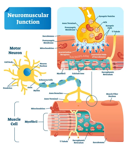
Neuromuscular Junction Vector Illustration Scheme. Labeled Cell Infographic
Vector, 7.8MB, 4000 × 4630 eps

Motor Neuron Connecting To Muscle Fiber, 3D Illustration. A Neuromuscular Junction Allows The Motor Neuron To Transmit A Signal To The Muscle Causing Contraction. It Is Affected By Toxins And Diseases
Image, 16.63MB, 7200 × 4050 jpg


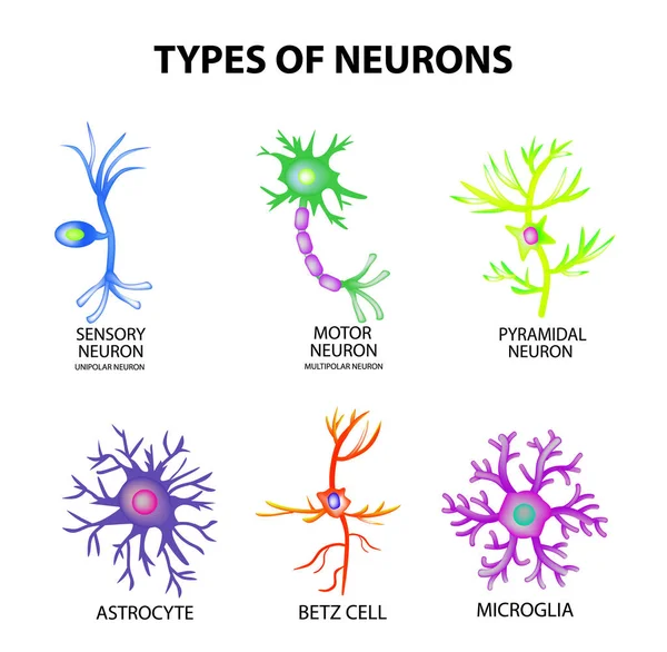
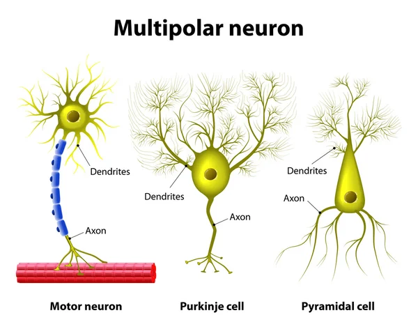
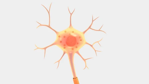
Types Of Neurons. Structure Sensory, Motor Neuron, Astrocyte, Pyromidal, Betz Cell, Microglia. Set. Infographics. Vector Illustration On Isolated Background
Vector, 3.45MB, 5000 × 4890 eps

Types Of Neurons. Structure Sensory, Motor Neuron, Astrocyte, Pyromidal, Betz Cell, Microglia. Set. Infographics. Vector
Vector, 3.45MB, 5000 × 4890 eps
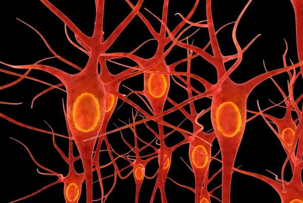
Neurons Of Dorsal Striatum, 3D Illustration. The Dorsal Striatum Is A Nucleus In The Basal Ganglia, Degrading Of Its Neurons Plays A Crucial Role In The Development Of Huntington's Disease
Image, 8.02MB, 7200 × 4050 jpg

Neurons Of Dorsal Striatum, 3D Illustration. Dorsal Striatum Is A Nucleus In The Basal Ganglia, Degrading Of Its Neurons Plays Crucial Role In Development Of Huntington's Disease
Image, 8.32MB, 7200 × 4050 jpg
Neurons Of Dorsal Striatum, 3D Illustration. Dorsal Striatum Is A Nucleus In The Basal Ganglia, Degrading Of Its Neurons Plays Crucial Role In Development Of Huntington's Disease
Image, 12.86MB, 7200 × 4050 jpg
Neurons Of Dorsal Striatum, 3D Illustration. The Dorsal Striatum Is A Nucleus In The Basal Ganglia, Degrading Of Its Neurons Plays A Crucial Role In The Development Of Huntington's Disease
Image, 12.53MB, 7200 × 4050 jpg

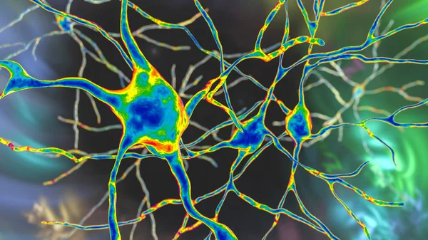
Neurons, Highly Detailed Brain Cells, Neural Network, 3D Illustration
Image, 12.08MB, 5120 × 5120 jpg


Motor Neuron Connecting To Muscle Fiber, 3D Illustration. A Neuromuscular Junction Allows The Motor Neuron To Transmit A Signal To The Muscle Causing Contraction. It Is Affected By Toxins And Diseases
Image, 15.53MB, 6075 × 4050 jpg
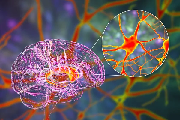
Caudate Nuclei In Human Brain And Its Neurons, 3D Illustration. The Caudate Nucleus Is A Component Of The Basal Ganglia, It Plays Role In Choreas, Neurodegenerative And Other Brain Diseases
Image, 16.77MB, 8157 × 5438 jpg

Motor Neuron Connecting To Muscle Fiber, 3D Illustration. A Neuromuscular Junction Allows The Motor Neuron To Transmit A Signal To The Muscle Causing Contraction. It Is Affected By Toxins And Diseases
Image, 16.17MB, 7200 × 4050 jpg
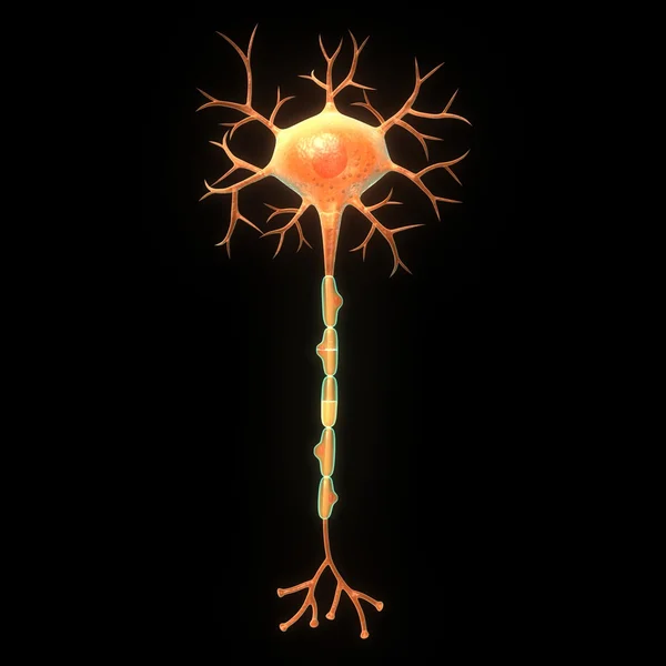
Demyelination Of Neuron, The Damage Of The Neuron Myelin Sheath Seen In Demyelinating Diseases, 3D Illustration. Multiple Sclerosis And Other Demyelinating Myelinoclastic And Leukodystrophic Diseases
Image, 17.84MB, 7200 × 4050 jpg
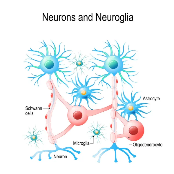
Neurons And Neuroglial Cells. Glial Cells Are Non-neuronal Cells In Brain. There Are Different Types Of Glial Cells: Oligodendrocyte, Microglia, Astrocytes And Schwann Cells. Vector Diagram For Educational, Medical, Biological And Science Use
Vector, 5.27MB, 4808 × 4808 eps

Demyelination Of Neuron, The Damage Of The Neuron Myelin Sheath Seen In Demyelinating Diseases, 3D Illustration. Multiple Sclerosis And Other Demyelinating Myelinoclastic And Leukodystrophic Diseases
Image, 24.2MB, 7200 × 4050 jpg

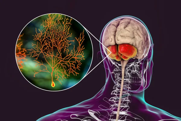
Human Brain With Highlighted Cerebellum And Close-up View Of Purkinje Neurons, One Of The Commonest Types Of Cells In Cerebellar Cortex, 3D Illustration
Image, 10.04MB, 8029 × 4517 jpg

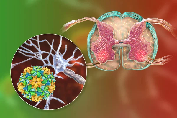
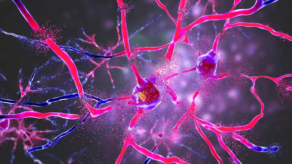
Mechanism Of Paralytic Polio Disease. Polio Viruses Affecting Motor Neurons Of Anterior Horn Of Spinal Cord, Conceptual 3D Illustration.
Image, 14.37MB, 8408 × 5605 jpg
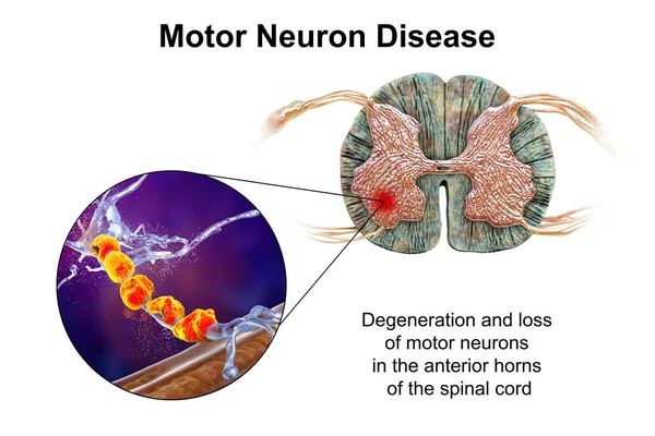
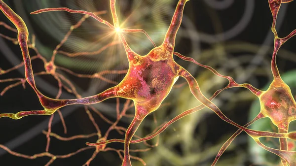
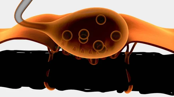
Motor Neuron Diseases, 3D Illustration Showing Degeneration Of Motor Neurons In Anterior Horns Of Spinal Cord. Amyotrophic Lateral Sclerosis And Other Motor Neuron Disorders
Image, 16.37MB, 10308 × 6872 jpg
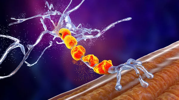
Degradation Of Motor Neurons, Conceptual 3D Illustration. Motor Neuron Diseases Are A Group Of Neurodegenerative Disorders Including Amyotrophic Lateral Sclerosis, Progressive Bulbar Palsy And Other
Image, 10.87MB, 7200 × 4050 jpg

World Day For The Fight Against Amyotrophic Lateral Sclerosis. Muscle And Neuron On White Background
Vector, 21.25MB, 8334 × 6251 eps

Caudate Nuclei In Human Brain And Its Neurons, 3D Illustration. The Caudate Nucleus Is A Component Of The Basal Ganglia, It Plays Role In Choreas, Neurodegenerative And Other Brain Diseases
Image, 12.8MB, 8157 × 5438 jpg
Human Brain With Highlighted Cerebellum And Close-up View Of Purkinje Neurons, One Of The Commonest Types Of Cells In Cerebellar Cortex, 3D Illustration
Image, 7.54MB, 6000 × 4000 jpg
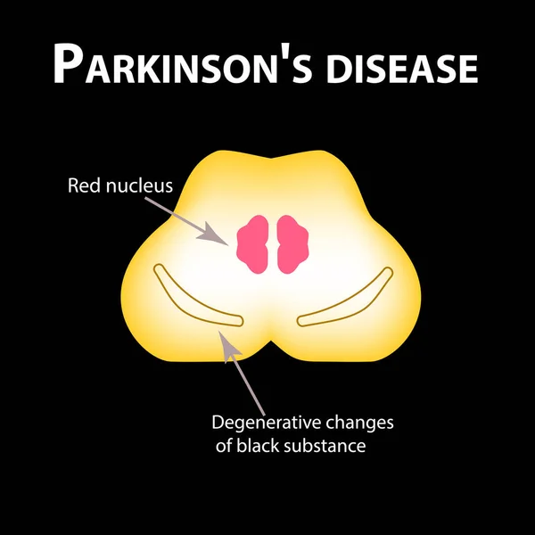
Parkinson's Disease. Degenerative Changes In The Brain Are A Black Substance. Vector Illustration On Black Background.
Vector, 0.89MB, 5000 × 5000 eps

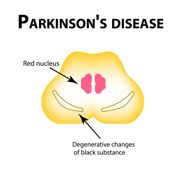
Parkinson's Disease. Degenerative Changes In The Brain Are A Black Substance. Vector Illustration On Isolated Background.
Vector, 0.89MB, 5000 × 5000 eps

Motor Neuron Diseases, 3D Illustration Showing Degeneration Of Motor Neurons In Anterior Horns Of Spinal Cord. Amyotrophic Lateral Sclerosis And Other Motor Neuron Disorders
Image, 14.98MB, 8408 × 5605 jpg


Demyelination Of Neuron, The Damage Of The Neuron Myelin Sheath Seen In Demyelinating Diseases, 3D Illustration. Multiple Sclerosis And Other Demyelinating Myelinoclastic And Leukodystrophic Diseases
Image, 20.55MB, 7200 × 4050 jpg
Demyelination Of Neuron, The Damage Of The Neuron Myelin Sheath Seen In Demyelinating Diseases, 3D Illustration. Multiple Sclerosis And Other Demyelinating Myelinoclastic And Leukodystrophic Diseases
Image, 12.46MB, 7200 × 4050 jpg
Motor Neuron Connecting To Muscle Fiber, 3D Illustration. A Neuromuscular Junction Allows The Motor Neuron To Transmit A Signal To The Muscle Causing Contraction. It Is Affected By Toxins And Diseases
Image, 10.6MB, 7200 × 4050 jpg

Motor Neuron Diseases, 3D Illustration Showing Degeneration Of Motor Neurons In Anterior Horns Of Spinal Cord. Amyotrophic Lateral Sclerosis And Other Motor Neuron Disorders
Image, 12.99MB, 10308 × 6872 jpg
Page 1 >> Next