
Stock image Mra Brain
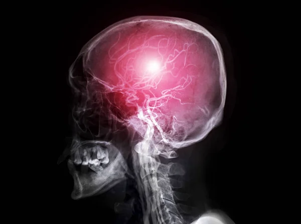
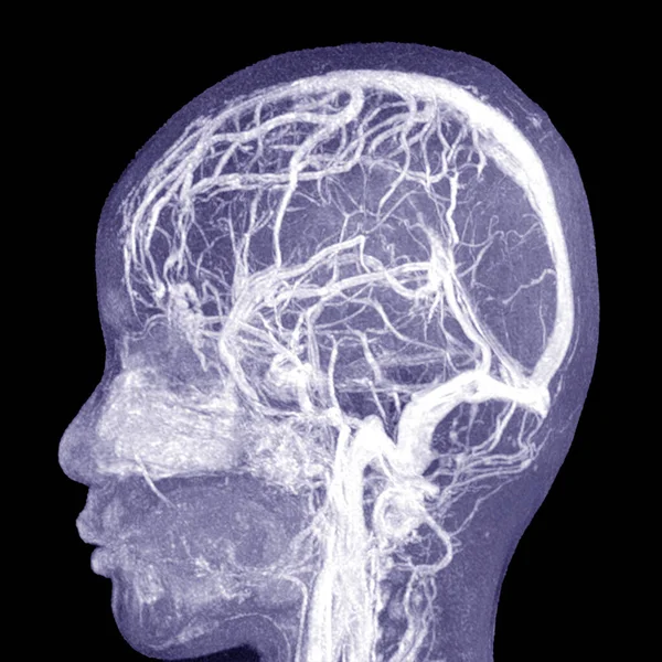
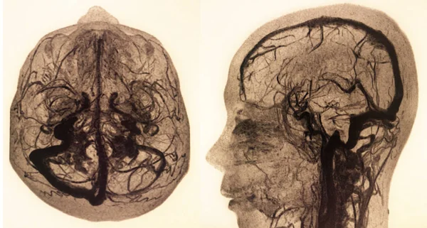
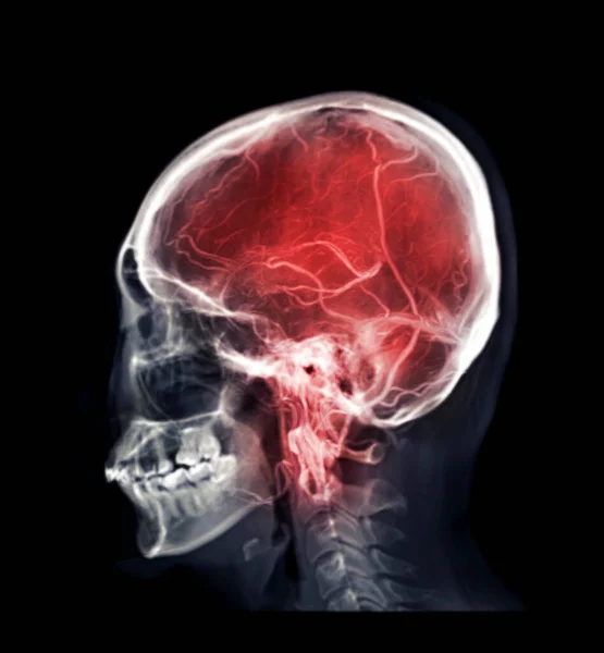
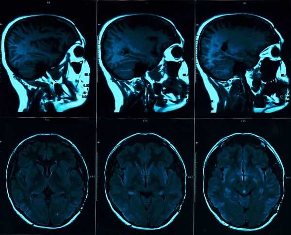
Skull X-ray Image Of Human Skull Lateral View Fusion With MRA Brain Image Showing Cerebral Artery Of Brain In Skull.
Image, 9.68MB, 7951 × 5928 jpg

Selective Focus Of Skull X-ray Image Of Human Skull Lateral View Fusion With MRA Brain Image .
Image, 9.99MB, 7951 × 5928 jpg
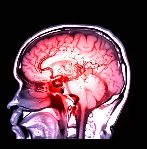
MRA Brain Or Magnetic Resonance Angiography Of Cerebral Artery In The Brain Sagittal View For Evaluate Them Stenosis And Stroke Disease.
Image, 3.59MB, 3083 × 3126 jpg
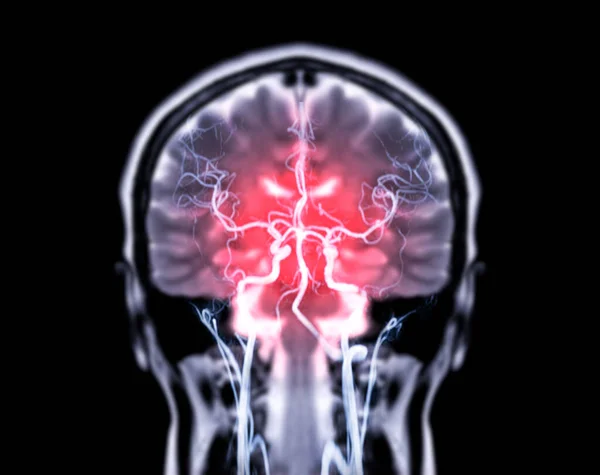
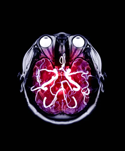
MRA Brain Or Magnetic Resonance Angiography ( MRA ) Of Cerebral Artery For Evaluate Them Aneurysm , Stenosis And Stroke Disease.
Image, 2.26MB, 3260 × 2757 jpg
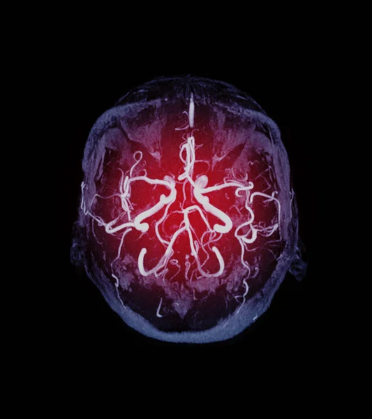
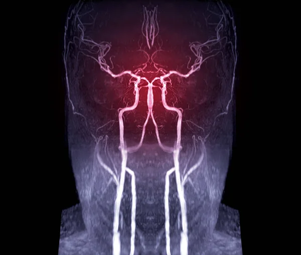
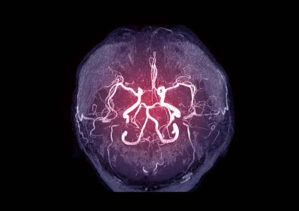
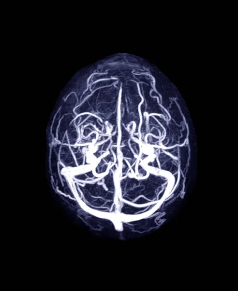
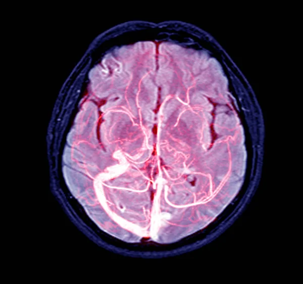
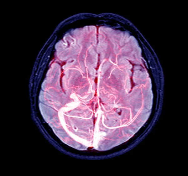
MRA Brain Axial MIP Veiw Showing Vessel In The Brain Name Is Circle Of Willis.
Image, 3MB, 4000 × 4500 jpg
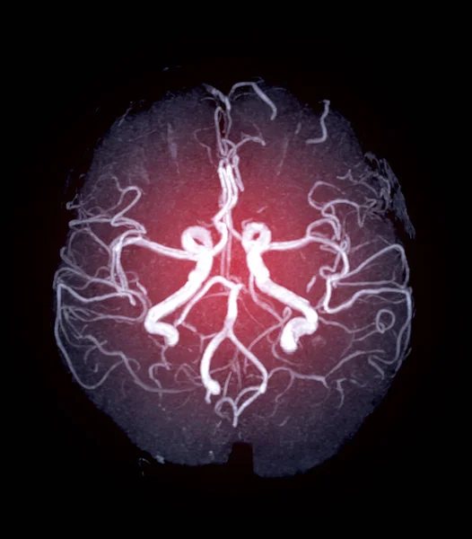
MRA Brain Or Magnetic Resonance Angiography (MRI) Of Vessel In The Brain Axial Mip View For Evaluate Them Stenosis , Occlusions, Aneurysms .
Image, 1.22MB, 2128 × 2424 jpg

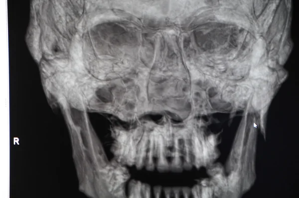
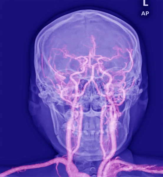
Skull X-ray Image Of Human Skull Ap Mix MRA Brain Image Showing Cerebral Artery Of Brain In Skull.
Image, 2.85MB, 3470 × 3848 jpg

Skull Image Fusion With MRI MRA Brain For Evaluate Them Stenosis And Stroke Disease.
Image, 3.51MB, 4491 × 2886 jpg

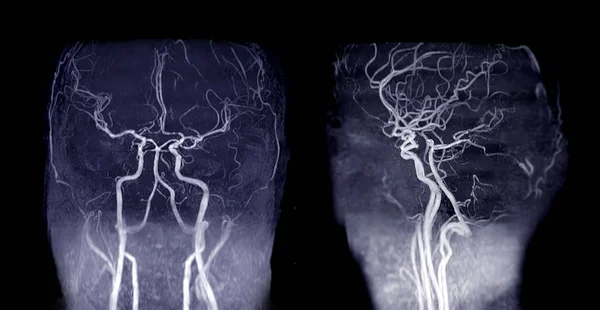
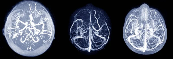
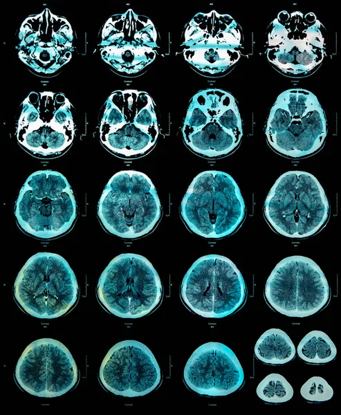
Collection Of MRA Brain Or Magnetic Resonance Angiography Image ( MRA ) Of Cerebral Artery In The Brain For Detect Stroke Disease.
Image, 2.38MB, 4780 × 3819 jpg

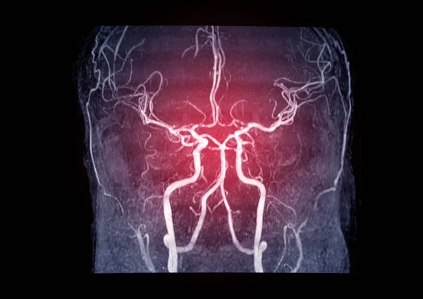
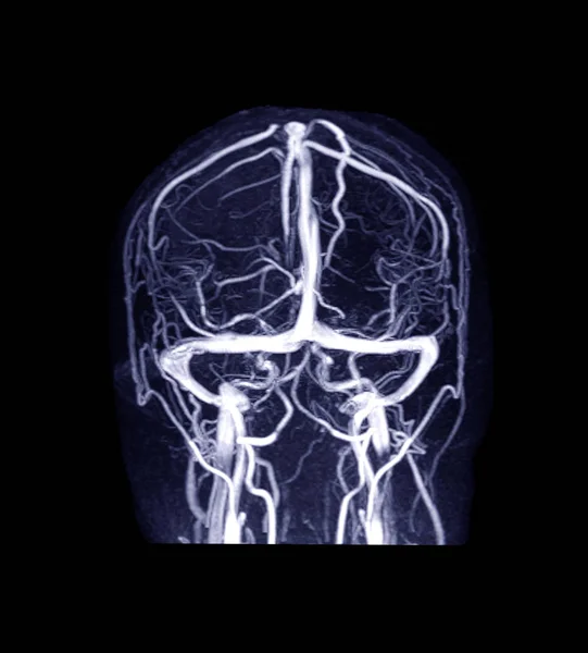
MRA Brain Coronal 3D MIP Veiw Showing Common Carotid Artery And Cerebral Artery .
Image, 1.53MB, 2706 × 3370 jpg
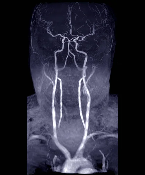
MRA Brain And Neck Or Magnetic Resonance Angiography ( MRA ) Of Cerebral Artery And Common Carotid Artery For Evaluate Them Stenosis And Stroke Disease.
Image, 3.19MB, 3392 × 4112 jpg
MRA Brain Or Magnetic Resonance Angiography ( MRA ) Of Cerebral Artery In The Brain For Evaluate Them Stenosis And Stroke Disease
Image, 1.46MB, 3392 × 2400 jpg
MRA Brain Or Magnetic Resonance Angiography ( MRA ) Of Cerebral Artery In The Brain For Evaluate Them Stenosis And Stroke Disease
Image, 1.97MB, 3392 × 2400 jpg
MRA Brain Or Magnetic Resonance Angiography ( MRA ) Of Cerebral Artery And Common Carotid Artery AP And Lateral View For Evaluate Them Stenosis And Stroke Disease.
Image, 3.83MB, 4980 × 2576 jpg
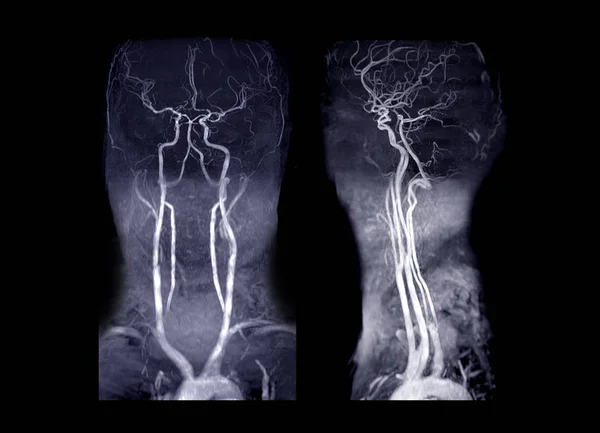
MRA Brain And Neck Or Magnetic Resonance Angiography ( MRA ) Of Cerebral Artery And Common Carotid Artery AP And Lateral View For Evaluate Them Stenosis And Stroke Disease.
Image, 4.76MB, 5696 × 4112 jpg
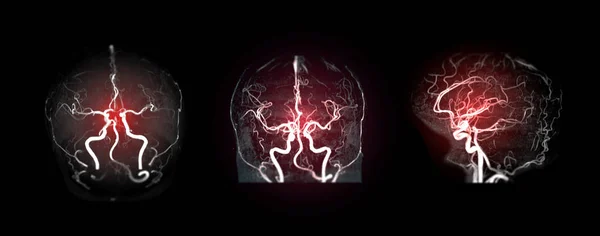
Collection Of MRA Brain Or Magnetic Resonance Angiography Image ( MRA ) Of Cerebral Artery In The Brain For Detect Stroke Disease.
Image, 5.35MB, 9698 × 3819 jpg

MRV Brain Or Magnetic Resonance Venography Of The Brain For Abnormalities In Venous Drainage Of The Brain
Image, 1.71MB, 2556 × 3112 jpg

MRI THE BRAIN.Moderate Perilesional Vasogenic Edema With 0.7 Cm Midline Shift To The Left Side.Medical Image Concept.
Image, 2.37MB, 3000 × 3000 jpg


MRV Brain Or Magnetic Resonance Venography Of The Brain For Abnormalities In Venous Drainage Of The Brain
Image, 1.96MB, 2700 × 2992 jpg
MRA AND MRV OF BRAIN Finding:Bilateral Territorial Muscles To The Cortex And Subcortical Of The Parietal And Low Hemispheres Cerebellar.
Image, 7.85MB, 4540 × 2430 jpg
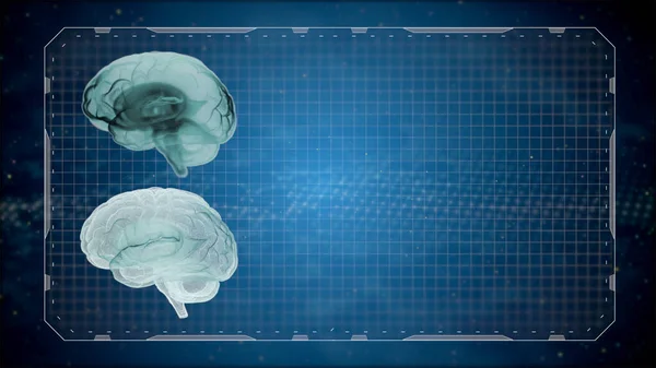
3d Render Computed Medical Tomography MRI Scan Of Human Brain Over Gridded Dark Background.
Image, 3.7MB, 5120 × 2880 jpg
MRA Brain Or Magnetic Resonance Angiography Image ( MRA ) Of Cerebral Artery In The Brain For Detect Stroke Disease.
Image, 2.31MB, 5332 × 3819 jpg
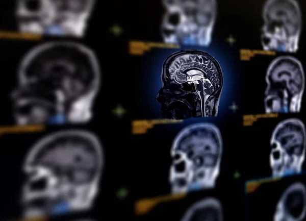
Selective Focus Of MRI Brain Sagittal Plane For Detect A Variety Of Conditions Of The Brain Such As Cysts, Tumors, Bleeding, Swelling, Developmental And Structural Abnormalities Or Infections .
Image, 7.51MB, 7584 × 5484 jpg
MRI OF THE BRAIN AND MRA & MRV OF THE BRAIN.Moderate Perilesional Vasogenic Edema With 0.7 Cm Midline Shift To The Left Side.
Image, 5.6MB, 8493 × 2920 jpg
Mix Skull Image And MRV Brain Or Magnetic Resonance Venography Of The Brain For Abnormalities In Venous Drainage Of The Brain
Image, 1.32MB, 2999 × 3240 jpg
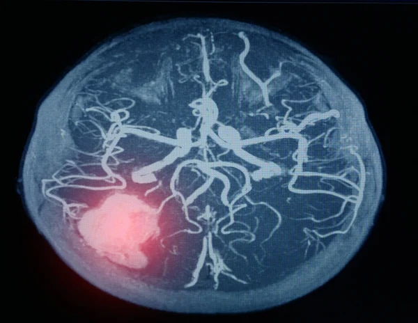
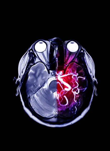
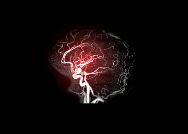
MRA Brain Or Magnetic Resonance Angiography Image ( MRA ) Of Cerebral Artery In The Hemorrhage In Brain.
Image, 5.8MB, 2970 × 2298 jpg

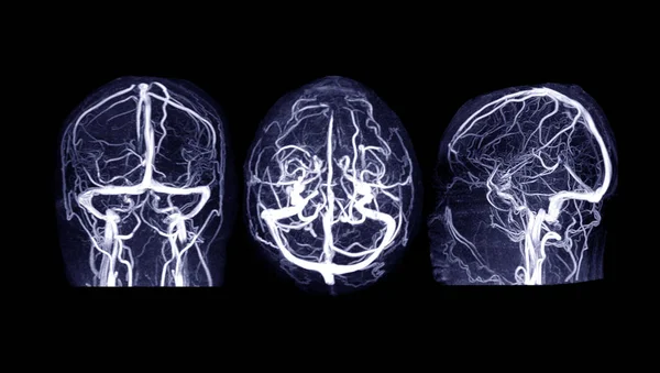
MRV Brain Or Magnetic Resonance Venography Of The Brain For Abnormalities In Venous Drainage Of The Brain
Image, 5.31MB, 6000 × 3400 jpg
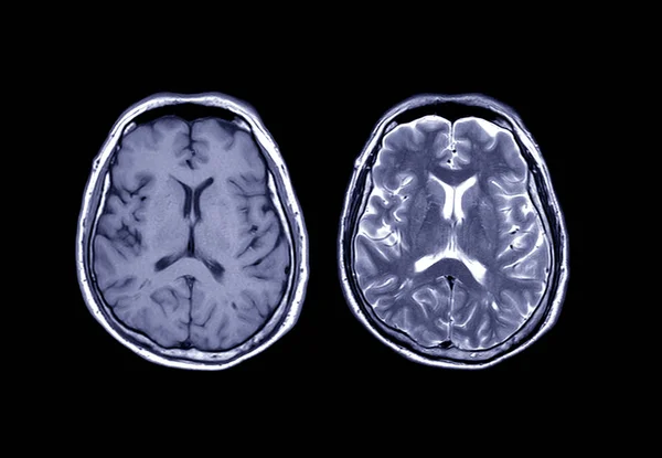
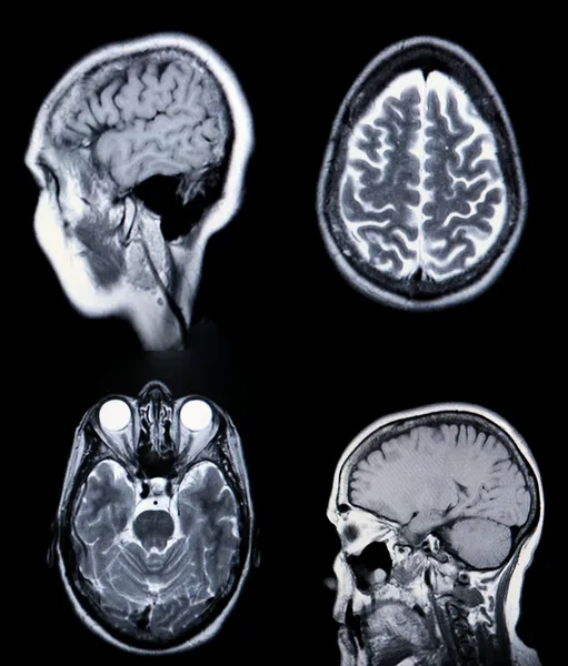
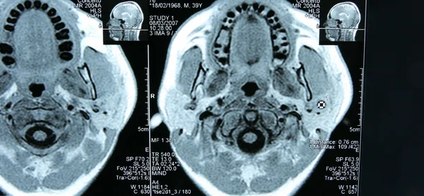
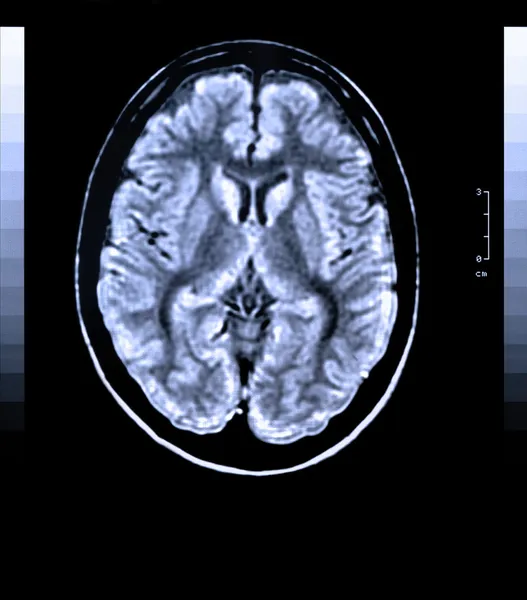
Comparison MRI Brain Axial T1 And T2 For Detect A Variety Of Conditions Of The Brain Such As Cysts, Tumors, Bleeding, Swelling, Developmental And Structural Abnormalities, Infections.
Image, 2.45MB, 4356 × 3016 jpg
Magnetic Resonance Scan Of The Brain With Skull. MRI Head Scan On Dark Background Blue Color. X-ray Medicine And Medication Concept
Image, 3.17MB, 2954 × 2388 jpg

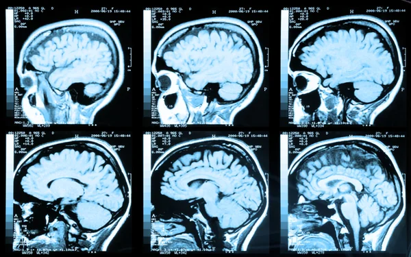
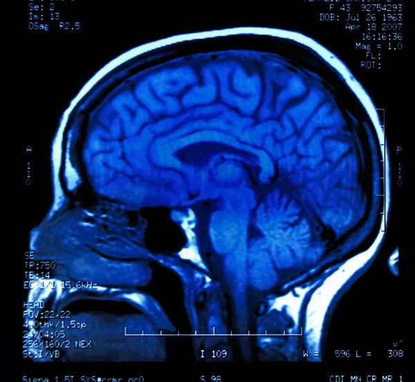
MRI Of The Brain Sagittal Plane For Detect A Variety Of Conditions Of The Brain Such As Cysts, Tumors, Bleeding, Swelling, Developmental And Structural Abnormalities, Infections.
Image, 4.62MB, 3104 × 4000 jpg

Selective Focus Of MRI Brain Sagittal Plane For Detect A Variety Of Conditions Of The Brain Such As Cysts, Tumors, Bleeding, Swelling, Developmental And Structural Abnormalities Or Infections .
Image, 6.81MB, 7056 × 5208 jpg
Page 1 >> Next