
Stock image Neoplasia

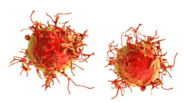
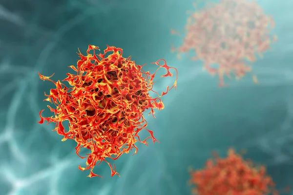
Cervical Cancer Cells, 3D Illustration. Malignant Tumor Of Cervix Uteri
Image, 13.47MB, 8617 × 4847 jpg
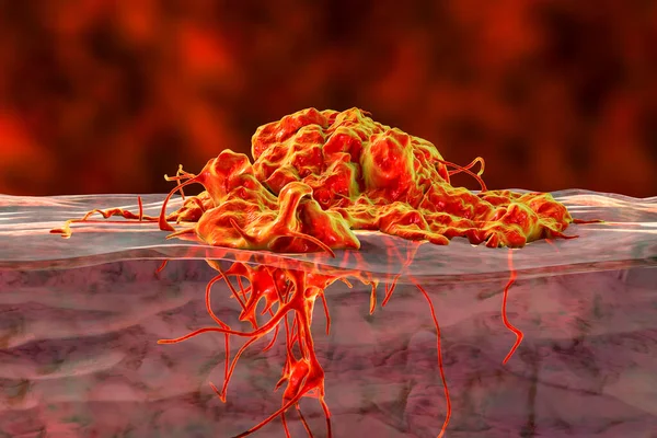
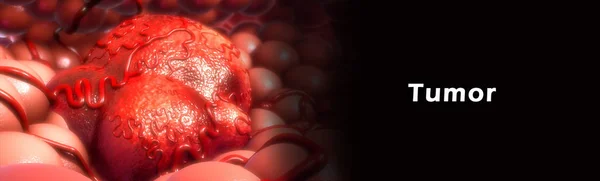
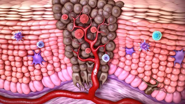
Invasive Cancer Growth, 3D Illustration Showing Tumor Invasion Into Underlined Tissue
Image, 9.15MB, 6000 × 4000 jpg

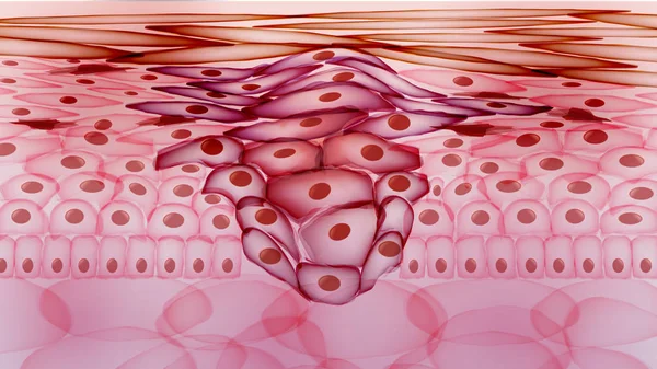
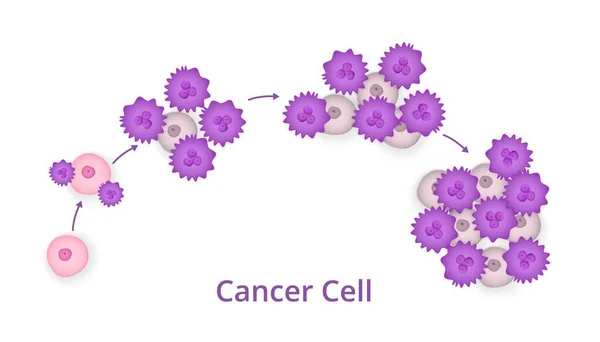
Cancer Cell Tumor, Medical Illustration. Neoplasm. Inflammation Caused By An Abnormal Growth Of Tissue, Whether Benign Or Malignant.
Image, 7.45MB, 4500 × 2500 jpg

The Scar On The Leg. Bone Fracture Or Tumor Removal. Recobvery. Isolated Medical On White Background
Image, 9.45MB, 5403 × 3602 jpg
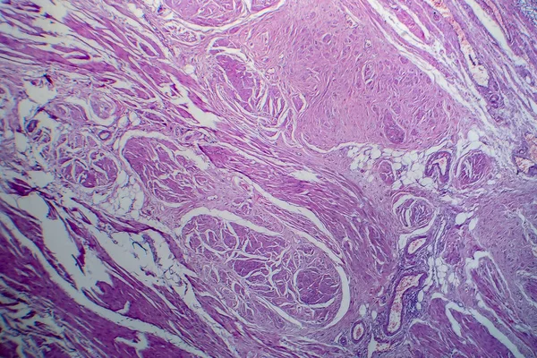
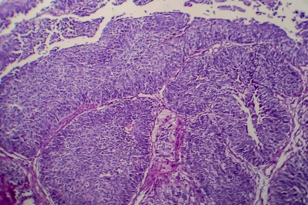
Bladder Transitional Cell Carcinoma, Light Micrograph, Photo Under Microscope
Image, 10.24MB, 4483 × 2989 jpg

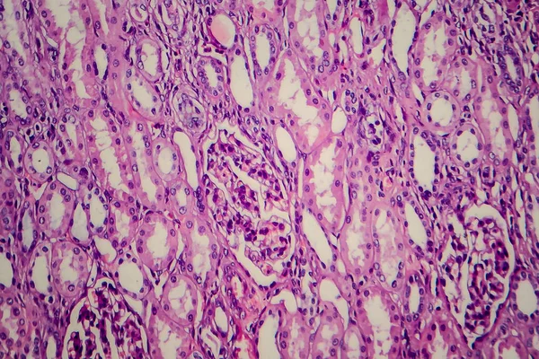
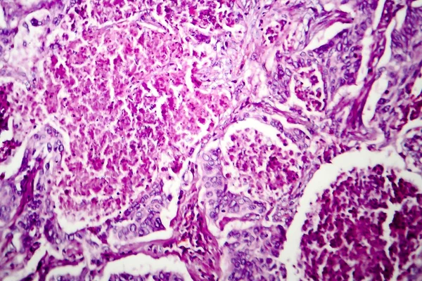
Wilms Tumor, Or Nephroblastoma, Light Micrograph, Photo Under Microscope
Image, 6.57MB, 3582 × 2388 jpg
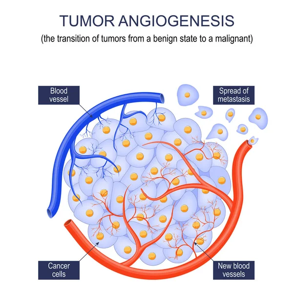
Tumor Angiogenesis. Transition Of Tumors From A Benign State To A Malignant. Tumor Grow. Cancer Cells And Spread Of Metastasis. Vector Poster
Vector, 6.7MB, 4444 × 4444 eps
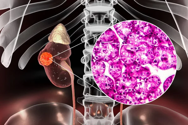
Kidney Cancer, Renal Cell Carcinoma, 3D Illustration And Light Micrograph
Image, 7.28MB, 4500 × 3000 jpg


Colorectal Cancer, Intestinal Carcinoma, Bowel Neoplasia, 3D Illustration Showing Malignant Tumor In Intestine
Image, 9.93MB, 7200 × 4050 jpg

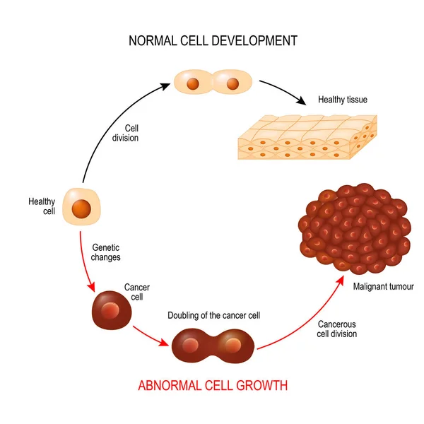
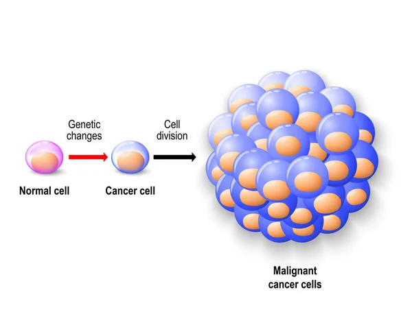
Cancer Cell And Normal Cell. Healthy Tissue And Malignant Tumour. Illustration Showing Cancer Disease Development. Vector Diagram For Your Design, Educational, Biological, Science And Medical Use
Vector, 2.66MB, 6102 × 6102 eps
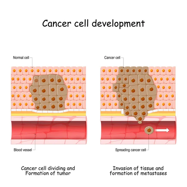
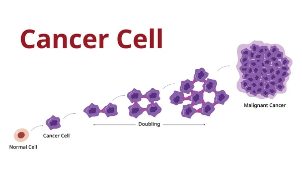
Cancer Development From Normal Cell To Formation Of Tumor, Spreading Cancer Cells In Blood Flow, Invasion Of Other Tissue, And Formation Of Metastases. Vector Illustration
Vector, 6.14MB, 4444 × 4444 eps

Cancer Development. Normal Cells Are Transformed Into Cancer. Carcinogenesis From Genetic Mutations In Healthy Cell To Malignant Cancer Cells. Mutagenesis, Oncogenesis Or Tumorigenesis. Tumor Formation. Vector Illustration.
Vector, 21.05MB, 6556 × 3000 eps

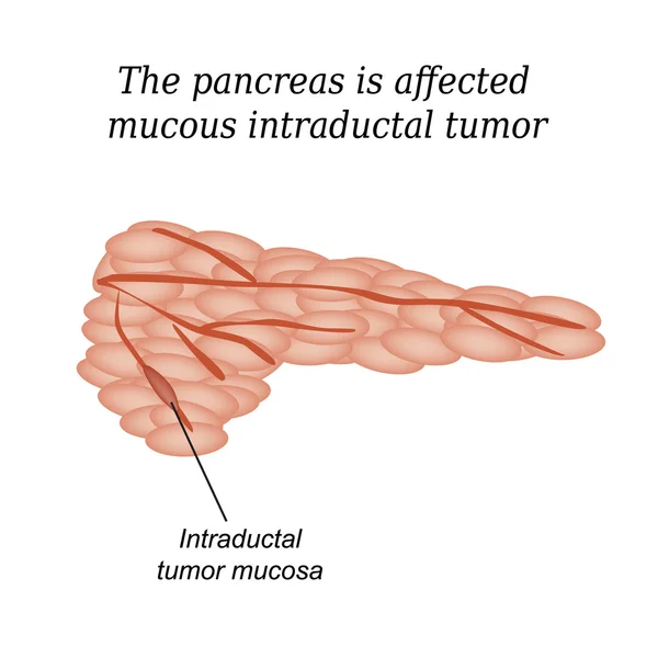
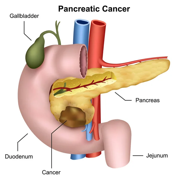
The Pancreas Is Affected Mucous Intraductal Tumor. Vector Illustration On Isolated Background
Vector, 4.21MB, 5000 × 5000 eps
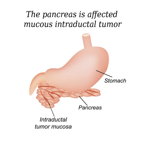
The Pancreas Is Affected Mucous Intraductal Tumor. Vector Illustration On Isolated Background
Vector, 3.73MB, 5000 × 5000 eps
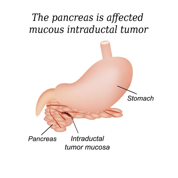
The Pancreas Is Affected Mucous Intraductal Tumor. Vector Illustration On Isolated Background
Vector, 3.37MB, 5000 × 5000 eps

Cervical Cancer Cells, 3D Illustration. Malignant Tumor Of Cervix Uteri
Image, 12.26MB, 7877 × 4431 jpg

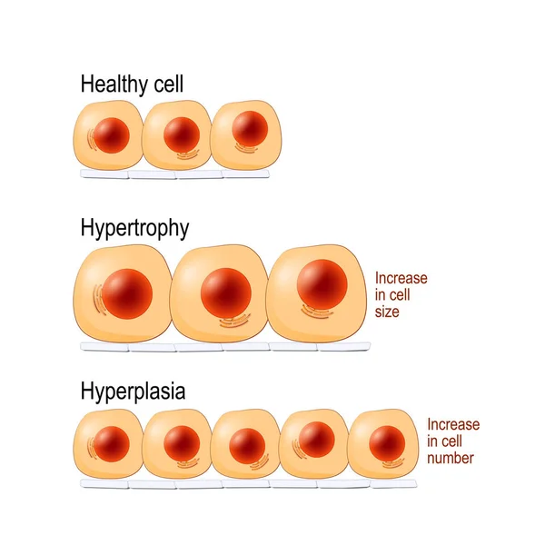
Normal Cells, Hypertrophy Is An Increase In Cell Size, Hyperplasia Results From An Increase In Cell Number. Different. Vector Diagram For Educational, Medical, Biological And Science Use
Vector, 7.14MB, 4462 × 4462 eps


The Scar On The Leg. Bone Fracture Or Tumor Removal. Recobvery. Isolated Medical On White Background
Image, 3.86MB, 4823 × 3005 jpg

Cervical Cancer Or Cervix Carcinoma. Development Stage. Vector Illustration
Vector, 10.45MB, 5000 × 3439 eps
3d Medical Illustration Of Skin Cancer: Squamous Cell Carcinoma, Basal Cell Cancer
Image, 2.15MB, 3840 × 2160 jpg

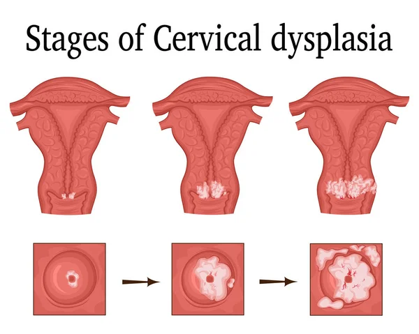
Cervical Dysplasia. Close-up Of A Cervix. Cervical Cancer. Cervical Intraepithelial Neoplasia Development From Normal Tissue And CIN I With Mild Dysplasia To Moderate Dysplasia And Squamous Cell Carcinoma. Vector Illustration Isolated On White Backgr
Vector, 9.8MB, 4444 × 4444 eps

A Human Body With Transparent Skin Showcasing Lung Cancer, 3D Illustration.
Image, 8.34MB, 6000 × 4000 jpg

A Pituitary Gland Tumor, 3D Medical Illustration Highlighting Its Location And Impact On Nearby Structures.
Image, 5.14MB, 6000 × 4000 jpg

A Human Body With Transparent Skin Showcasing Lung Cancer, 3D Illustration Complemented By A Light Micrograph Of The Lung Adenocarcinoma.
Image, 10.23MB, 6000 × 4000 jpg
Bladder Transitional Cell Carcinoma, Light Micrograph, Photo Under Microscope
Image, 10.33MB, 4483 × 2989 jpg

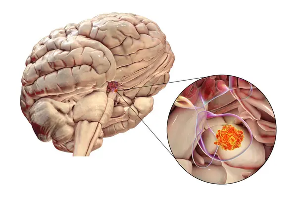
A Pituitary Gland Tumor, 3D Medical Illustration Highlighting Its Location And Impact On Nearby Structures.
Image, 10.43MB, 9000 × 6000 jpg
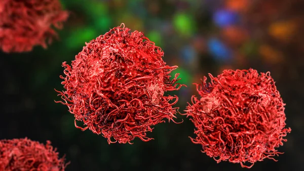
Stomach Cancer Cells, 3D Illustration Showing Morphology Of Cancerous Cells
Image, 16.21MB, 8347 × 4695 jpg
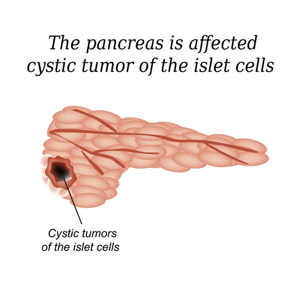
The Pancreas Is Affected Cystic Tumor Of The Islet Cells. Vector Illustration On Isolated Background
Vector, 4.16MB, 5000 × 5000 eps
Page 1 >> Next