
Stock image Neoplasia page 2

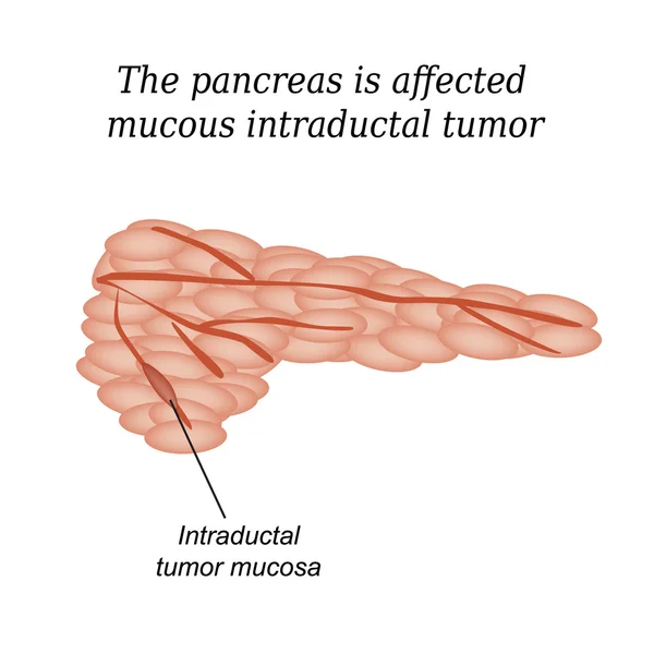
The Pancreas Is Affected Mucous Intraductal Tumor. Vector Illustration On Isolated Background
Vector, 4.21MB, 5000 × 5000 eps
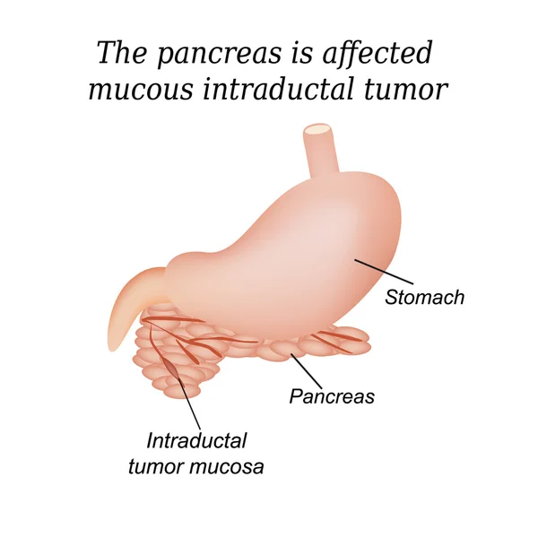
The Pancreas Is Affected Mucous Intraductal Tumor. Vector Illustration On Isolated Background
Vector, 3.73MB, 5000 × 5000 eps
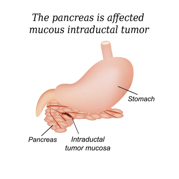
The Pancreas Is Affected Mucous Intraductal Tumor. Vector Illustration On Isolated Background
Vector, 3.37MB, 5000 × 5000 eps

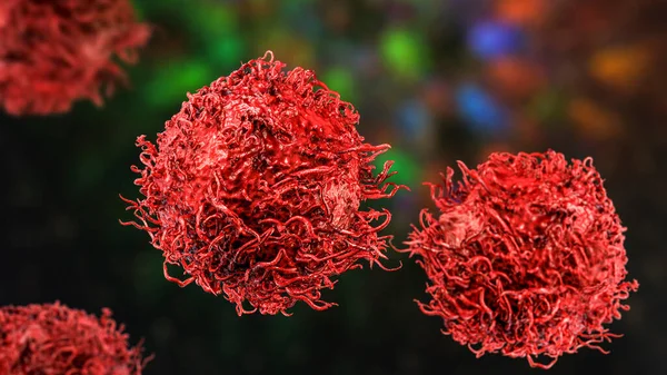
Cervical Cancer Cells, 3D Illustration. Malignant Tumor Of Cervix Uteri
Image, 12.26MB, 7877 × 4431 jpg

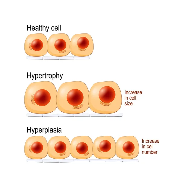
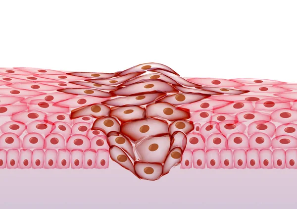
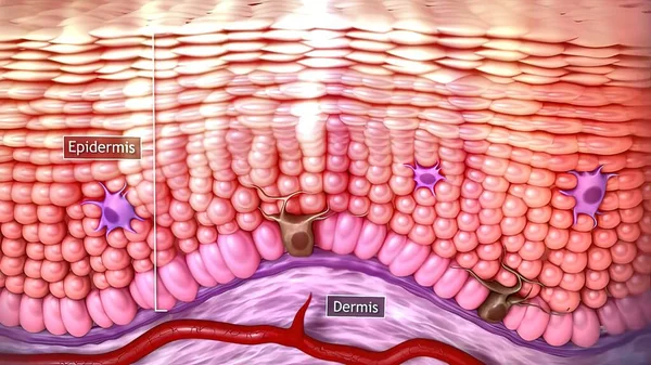
Normal Cells, Hypertrophy Is An Increase In Cell Size, Hyperplasia Results From An Increase In Cell Number. Different. Vector Diagram For Educational, Medical, Biological And Science Use
Vector, 7.14MB, 4462 × 4462 eps


The Scar On The Leg. Bone Fracture Or Tumor Removal. Recobvery. Isolated Medical On White Background
Image, 3.86MB, 4823 × 3005 jpg

Cervical Cancer Or Cervix Carcinoma. Development Stage. Vector Illustration
Vector, 10.45MB, 5000 × 3439 eps
3d Medical Illustration Of Skin Cancer: Squamous Cell Carcinoma, Basal Cell Cancer
Image, 2.15MB, 3840 × 2160 jpg

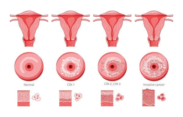
Cervical Dysplasia. Close-up Of A Cervix. Cervical Cancer. Cervical Intraepithelial Neoplasia Development From Normal Tissue And CIN I With Mild Dysplasia To Moderate Dysplasia And Squamous Cell Carcinoma. Vector Illustration Isolated On White Backgr
Vector, 9.8MB, 4444 × 4444 eps
Set Of Cervical Invasive Cancer Intraepithelial Neoplasia Dysplasia 1 2,3 And Normal Stage Cell Morphology On Pap Smear Test Infographic Female Reproductive System Carcinoma View. Medical Flat Style
Vector, 10.89MB, 9308 × 6000 eps


A Human Body With Transparent Skin Showcasing Lung Cancer, 3D Illustration Complemented By A Light Micrograph Of The Lung Adenocarcinoma.
Image, 10.23MB, 6000 × 4000 jpg

Cancer Evolution From Mutated Cell And Hyperplasia, To Dysplasia And Malignant Tumor. Spread Cancer Cells To Other Tissues. Vector Diagram
Vector, 3.51MB, 5164 × 3000 eps
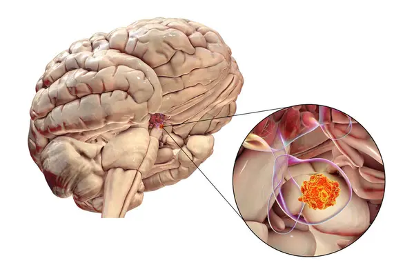
A Pituitary Gland Tumor, 3D Medical Illustration Highlighting Its Location And Impact On Nearby Structures.
Image, 10.43MB, 9000 × 6000 jpg

A Human Body With Transparent Skin Showcasing Lung Cancer, 3D Illustration.
Image, 8.34MB, 6000 × 4000 jpg
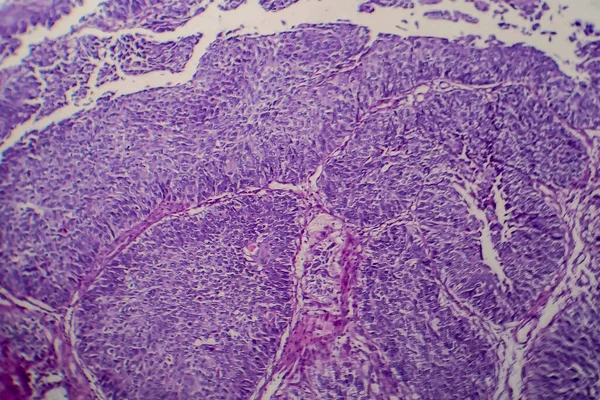
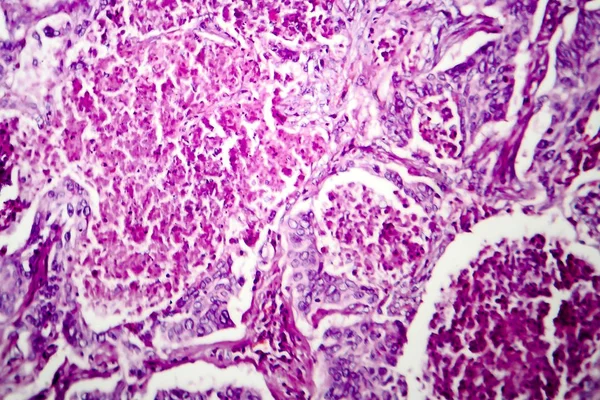
Bladder Transitional Cell Carcinoma, Light Micrograph, Photo Under Microscope
Image, 10.33MB, 4483 × 2989 jpg

A Pituitary Gland Tumor, 3D Medical Illustration Highlighting Its Location And Impact On Nearby Structures.
Image, 5.14MB, 6000 × 4000 jpg

Cervical Intraepithelial Neoplasia. Cervical Dysplasia. CIN. Pre-cancerous Lesion. Close-up Of A Cervix. Cancer Development From Normal Cell To Precancerous Transformation Of Cells Of The Cervix. Vector. Schematic Diagram. Detailed Poster.
Vector, 6.93MB, 4444 × 4444 eps
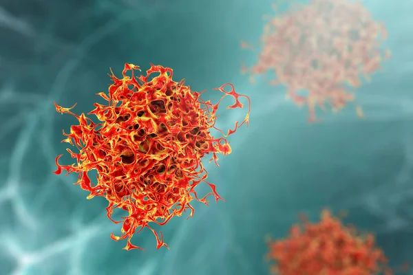
Stomach Cancer Cells, 3D Illustration Showing Morphology Of Cancerous Cells
Image, 16.21MB, 8347 × 4695 jpg
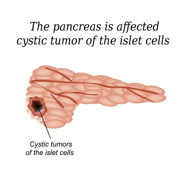
The Pancreas Is Affected Cystic Tumor Of The Islet Cells. Vector Illustration On Isolated Background
Vector, 4.16MB, 5000 × 5000 eps

CT Scan Of The Abdomen Of A Patient With Carcinoma Of The Head Of Pancreas.
Image, 12.33MB, 6096 × 3896 jpg

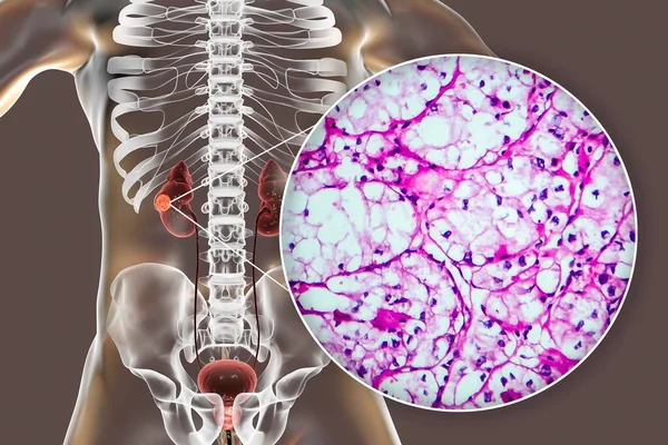
Kidney Cancer, Renal Cell Carcinoma, 3D Illustration And Light Micrograph
Image, 8.61MB, 5334 × 3000 jpg
Kidney Cancer, Renal Cell Carcinoma, 3D Illustration And Light Micrograph
Image, 6.41MB, 4500 × 3000 jpg
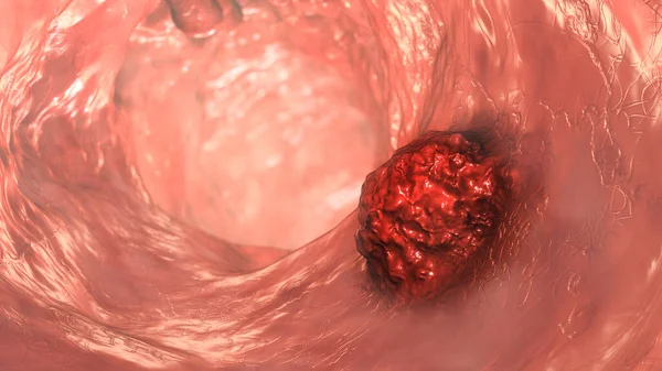
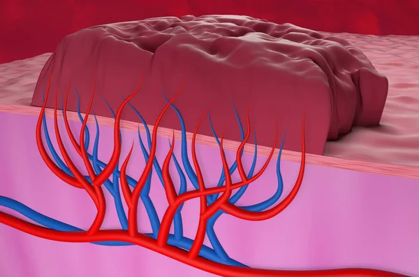
Colorectal Cancer, Intestinal Carcinoma, Bowel Neoplasia, 3D Illustration Showing Malignant Tumor In Intestine
Image, 9.14MB, 7200 × 4050 jpg


Cervical Dysplasia. Difference In Covering Epithelium Affected Of Cervical Intraepithelial Neoplasia. Cross Section Of Epithelium. Close-up Of A Normal Squamous Cell And Abnormal Cells. Cervical Cancer Evolution. Vector Illustration
Vector, 8.97MB, 6161 × 3000 eps
3d Medical Illustration Of Skin Cancer: Squamous Cell Carcinoma, Basal Cell Cancer
Image, 2.02MB, 3840 × 2160 jpg

Doctor Makes A Patient Patient A Thyroid Biopsy On Suspicion Of Oncology, Thyroid Node, Close-up, Medic, Endocrinology
Image, 8.63MB, 5184 × 3246 jpg
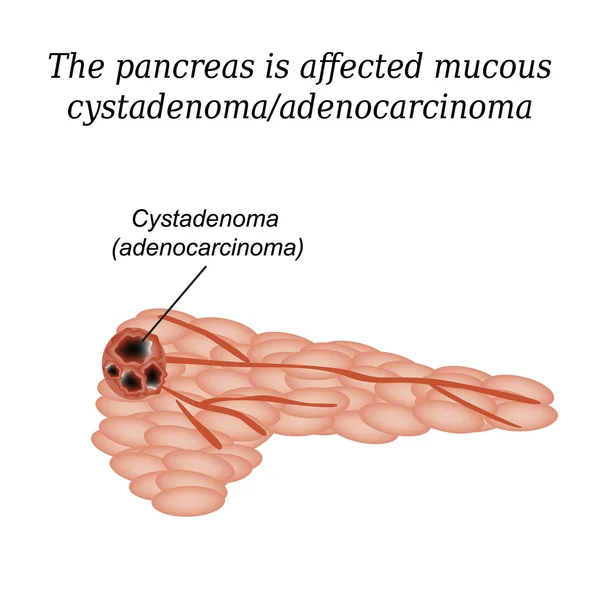
Pancreas Mucous Cystadenoma, Adenocarcinoma. Vector Illustration On Isolated Background
Vector, 4.46MB, 5000 × 5000 eps

The Doctor Makes The Woman Puncture The Thyroid Gland To Clarify The Diagnosis, Cancer, Copy Space
Image, 8.5MB, 5046 × 3456 jpg
Colorectal Cancer, Intestinal Carcinoma, Bowel Neoplasia, 3D Illustration Showing Malignant Tumor In Intestine
Image, 10.49MB, 7200 × 4050 jpg

Kidney Cancer, Renal Cell Carcinoma, 3D Illustration And Light Micrograph
Image, 10.16MB, 6121 × 4080 jpg
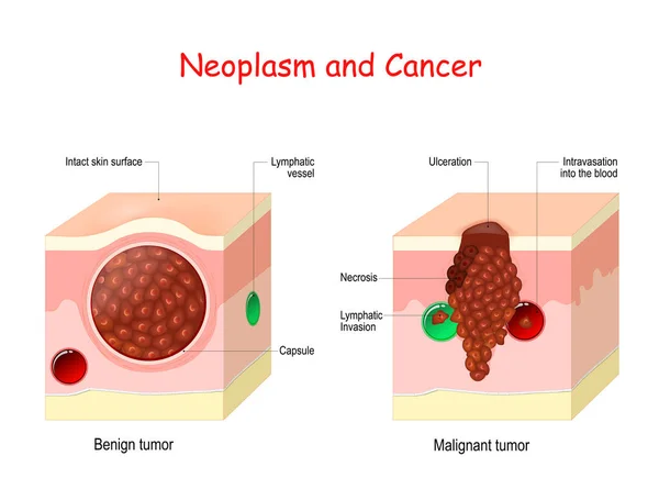
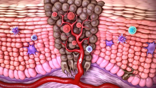
Cancer And Neoplasm. Comparison And Difference Between Malignant And Benign Tumor. Benign Tumor Has A Capsule. Cells Of Malignant Tumor Have Necrosis, Intravasation Into The Blood Vessel, And Lymphatic Invasion.
Vector, 12.6MB, 5000 × 3715 eps

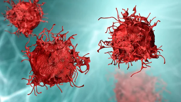
Doctor Holds Two Vials Of Idarubicina And Oncaspar To Inject, Medicine Used In Acute Lymphatic Leukemia Disease
Image, 7.09MB, 5184 × 3456 jpg
Previous << Page 2 >> Next