
Stock image Neoplasia page 3
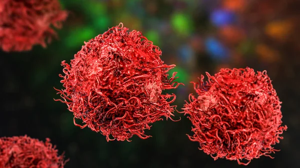
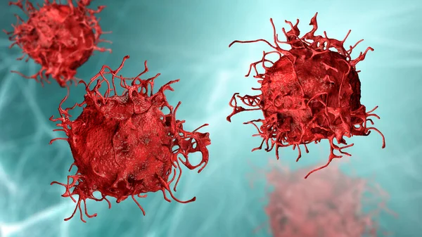
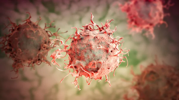
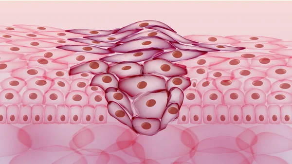
Stomach Cancer Cells, 3D Illustration Showing Morphology Of Cancerous Cells
Image, 16.21MB, 8347 × 4695 jpg
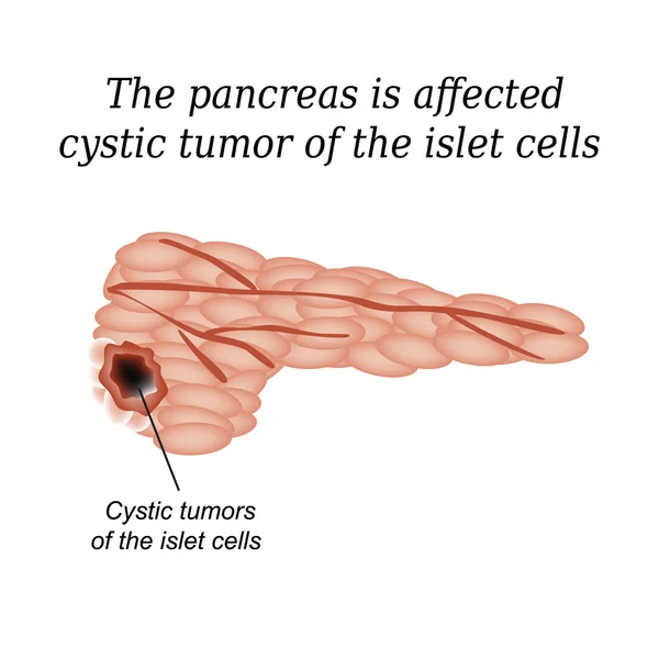
The Pancreas Is Affected Cystic Tumor Of The Islet Cells. Vector Illustration On Isolated Background
Vector, 4.16MB, 5000 × 5000 eps

CT Scan Of The Abdomen Of A Patient With Carcinoma Of The Head Of Pancreas.
Image, 12.33MB, 6096 × 3896 jpg

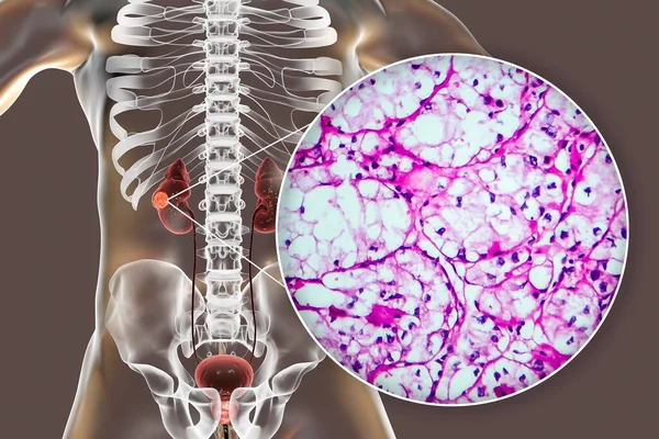
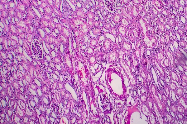
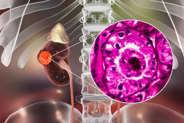
Kidney Cancer, Renal Cell Carcinoma, 3D Illustration And Light Micrograph
Image, 8.61MB, 5334 × 3000 jpg
Kidney Cancer, Renal Cell Carcinoma, 3D Illustration And Light Micrograph
Image, 6.41MB, 4500 × 3000 jpg
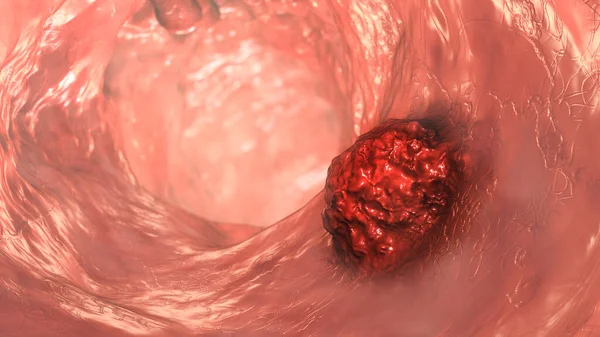
Colorectal Cancer, Intestinal Carcinoma, Bowel Neoplasia, 3D Illustration Showing Malignant Tumor In Intestine
Image, 9.14MB, 7200 × 4050 jpg


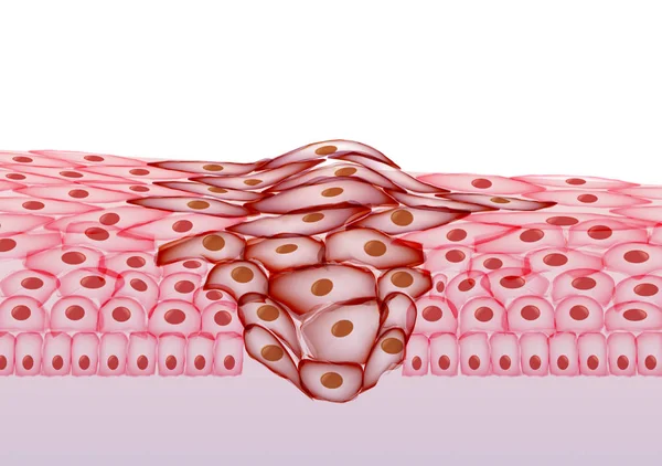
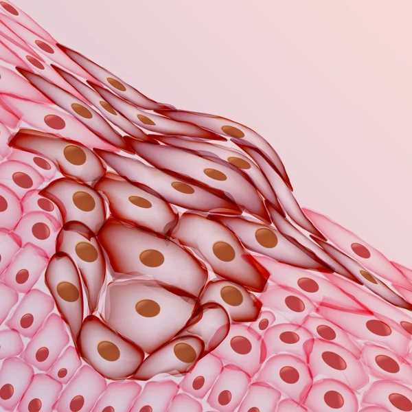
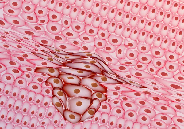
Cervical Dysplasia. Difference In Covering Epithelium Affected Of Cervical Intraepithelial Neoplasia. Cross Section Of Epithelium. Close-up Of A Normal Squamous Cell And Abnormal Cells. Cervical Cancer Evolution. Vector Illustration
Vector, 8.97MB, 6161 × 3000 eps
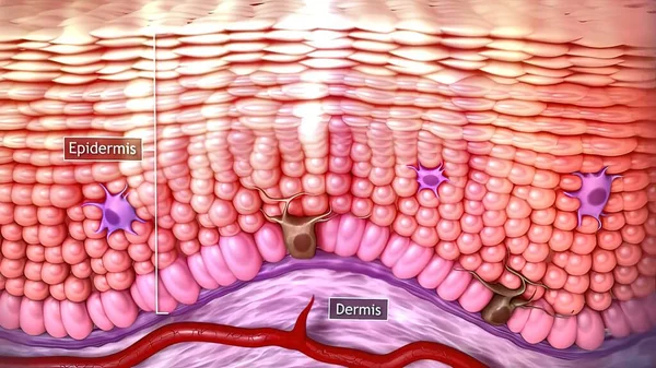
3d Medical Illustration Of Skin Cancer: Squamous Cell Carcinoma, Basal Cell Cancer
Image, 2.02MB, 3840 × 2160 jpg

Doctor Makes A Patient Patient A Thyroid Biopsy On Suspicion Of Oncology, Thyroid Node, Close-up, Medic, Endocrinology
Image, 8.63MB, 5184 × 3246 jpg
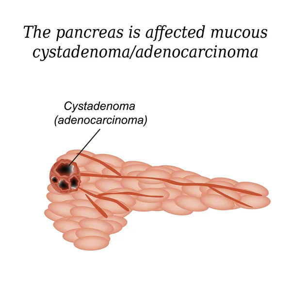
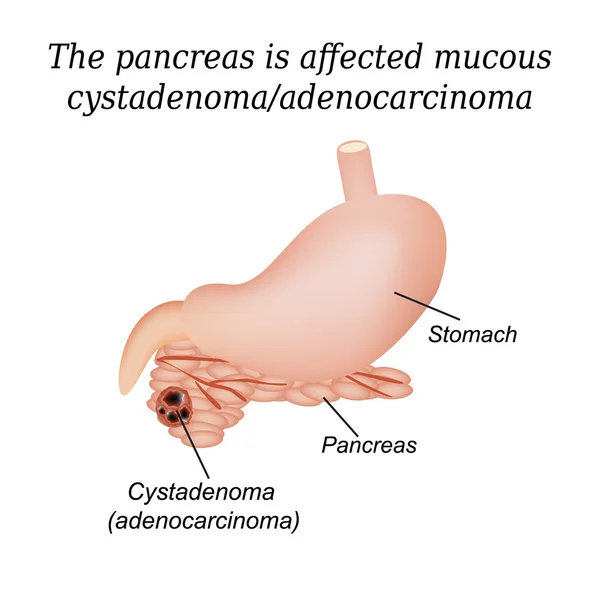
Pancreas Mucous Cystadenoma, Adenocarcinoma. Vector Illustration On Isolated Background
Vector, 4.46MB, 5000 × 5000 eps

The Doctor Makes The Woman Puncture The Thyroid Gland To Clarify The Diagnosis, Cancer, Copy Space
Image, 8.5MB, 5046 × 3456 jpg
Colorectal Cancer, Intestinal Carcinoma, Bowel Neoplasia, 3D Illustration Showing Malignant Tumor In Intestine
Image, 10.49MB, 7200 × 4050 jpg

Kidney Cancer, Renal Cell Carcinoma, 3D Illustration And Light Micrograph
Image, 10.16MB, 6121 × 4080 jpg
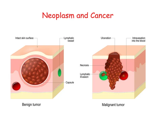
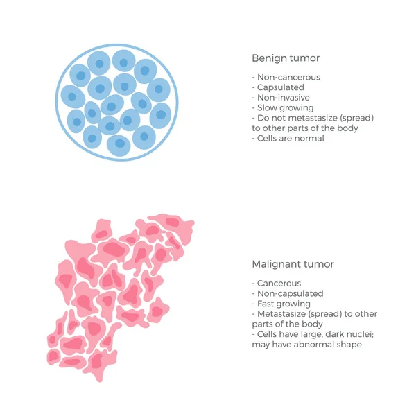
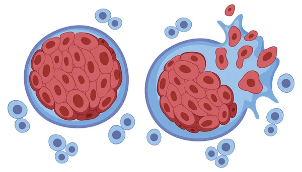
Cancer And Neoplasm. Comparison And Difference Between Malignant And Benign Tumor. Benign Tumor Has A Capsule. Cells Of Malignant Tumor Have Necrosis, Intravasation Into The Blood Vessel, And Lymphatic Invasion.
Vector, 12.6MB, 5000 × 3715 eps

Doctor Holds Two Vials Of Idarubicina And Oncaspar To Inject, Medicine Used In Acute Lymphatic Leukemia Disease
Image, 7.09MB, 5184 × 3456 jpg

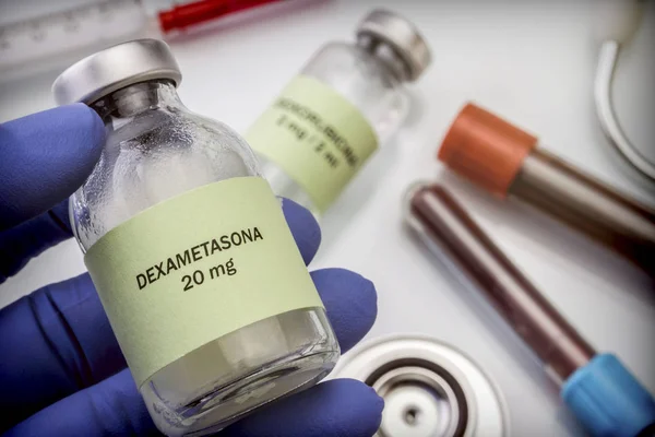
Doctor Holds Vial Of Dexamethasone In An Operating Theater, Conceptual Image
Image, 5.68MB, 5184 × 3601 jpg
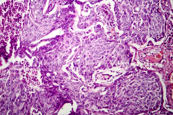
Wilms Tumor, Or Nephroblastoma, Light Micrograph, Photo Under Microscope
Image, 8.15MB, 3582 × 2388 jpg
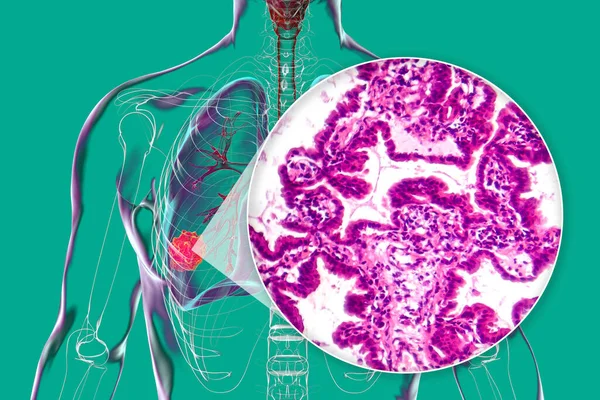
A Human Body With Transparent Skin Showcasing Lung Cancer, 3D Illustration Complemented By A Light Micrograph Of The Lung Adenocarcinoma.
Image, 10.12MB, 5999 × 4000 jpg

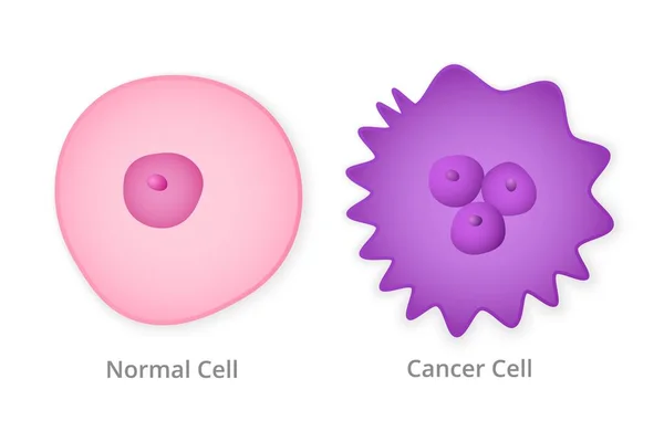
Malignant Neoplasm. Cancer And Normal Cells. Comparison And Difference Between Healthy Tissue And Tumor. Details About Chromatin, Nucleus And Cytoplasm
Vector, 1.59MB, 4444 × 4444 eps
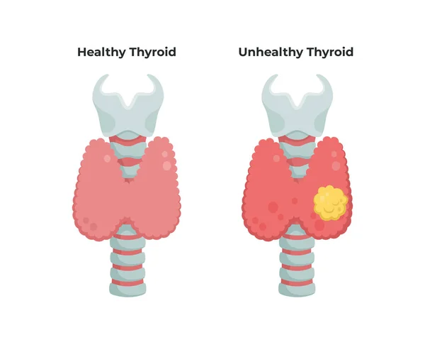
Healthy Thyroid Gland And Unhealthy Thyroid With Inflammation And Lump, Thyroid Cancer Concept, Flat Illustration Isolated On White Background. Medical Infographic Elements.
Vector, 5.49MB, 6946 × 5557 eps
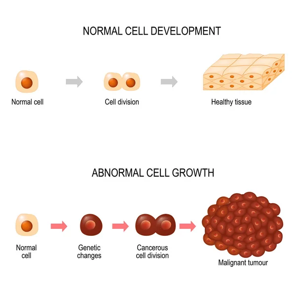
Cancer Cells. Illustration Showing Cancer Disease Development. Healthy Tissue And Malignant Tumour. Vector Diagram For Your Design, Educational, Biological, Science And Medical Use
Vector, 2.5MB, 5207 × 5206 eps

Cervical Cancer Cells, 3D Illustration. Malignant Tumor Of Cervix Uteri
Image, 13.19MB, 7877 × 4431 jpg

Uterine Cancer. Cross Section Of A Human Uterus With Endometrial Tumor. Female Reproductive System. Vector Illustration Like X-ray Image. Reproductive Health.
Vector, 2.36MB, 4444 × 4444 eps
Pancreas Mucous Cystadenoma, Adenocarcinoma. Vector Illustration On Isolated Background
Vector, 4.26MB, 5000 × 5000 eps
Kidney Cancer, Renal Cell Carcinoma, 3D Illustration And Light Micrograph
Image, 7.09MB, 4500 × 3000 jpg

Cervical Cancer Cells, 3D Illustration. Malignant Tumor Of Cervix Uteri
Image, 6.4MB, 7200 × 4050 jpg

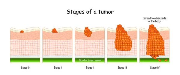
Stages Of Cancer. Classification Of Malignant Tumours (from 0 To 4). System That Is Most Commonly Used For The Staging Process Of Cancer
Vector, 8.55MB, 7000 × 2936 eps

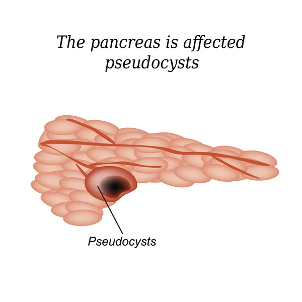
Pancreas Pseudocyst Affected. Vector Illustration On Isolated Background
Vector, 4.12MB, 5000 × 5000 eps
Previous << Page 3 >> Next