
Stock image Neuromuscular Junction
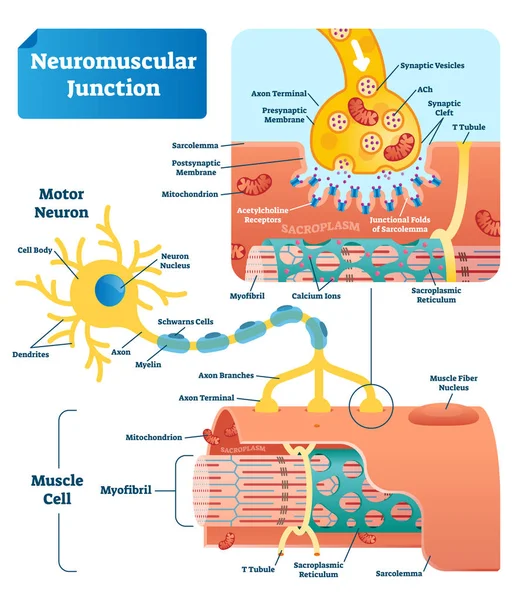
Neuromuscular Junction Vector Illustration Scheme. Labeled Cell Infographic
Vector, 7.8MB, 4000 × 4630 eps

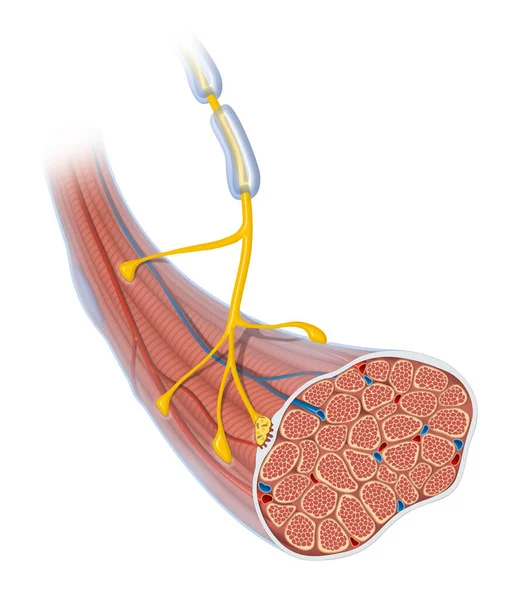
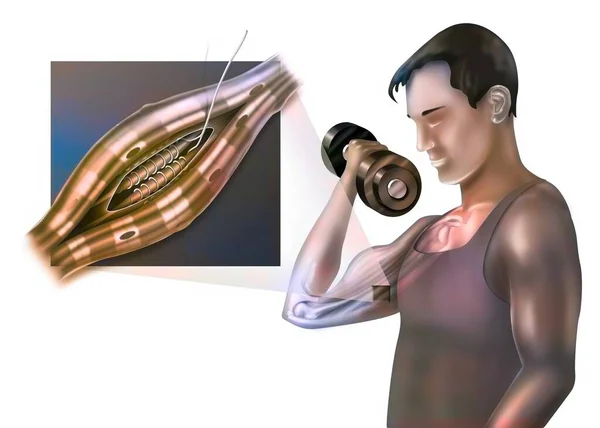
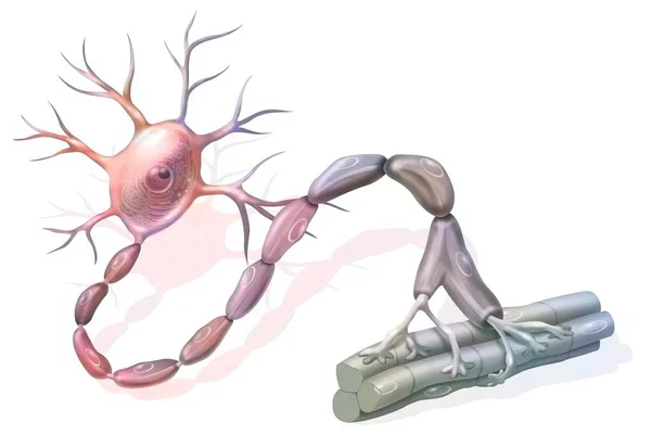
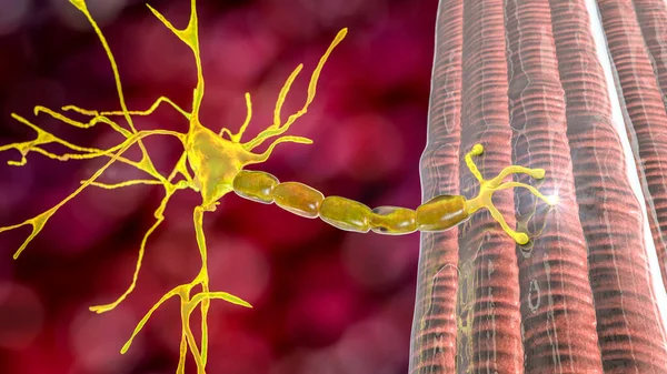
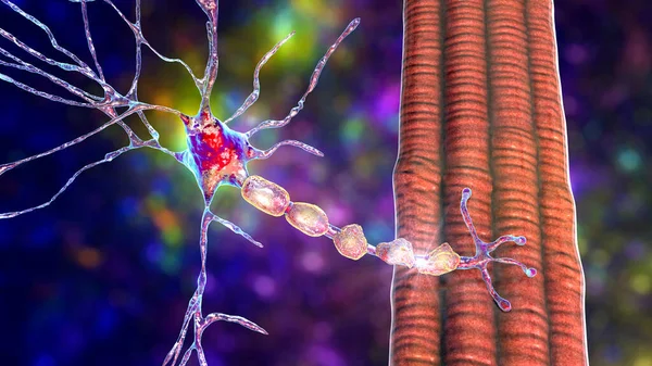
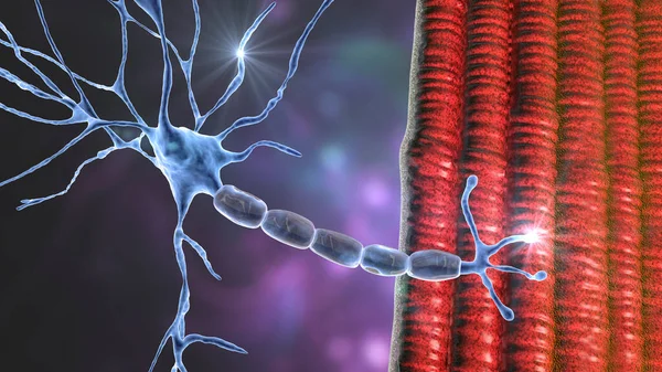
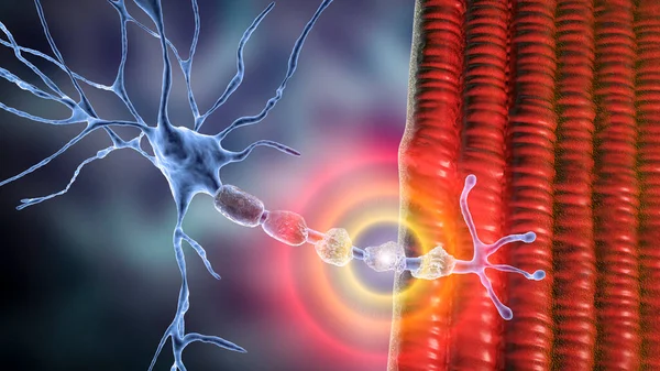
Motor Neuron Connecting To Muscle Fiber, 3D Illustration. A Neuromuscular Junction Allows The Motor Neuron To Transmit A Signal To The Muscle Causing Contraction. It Is Affected By Toxins And Diseases
Image, 16.63MB, 7200 × 4050 jpg
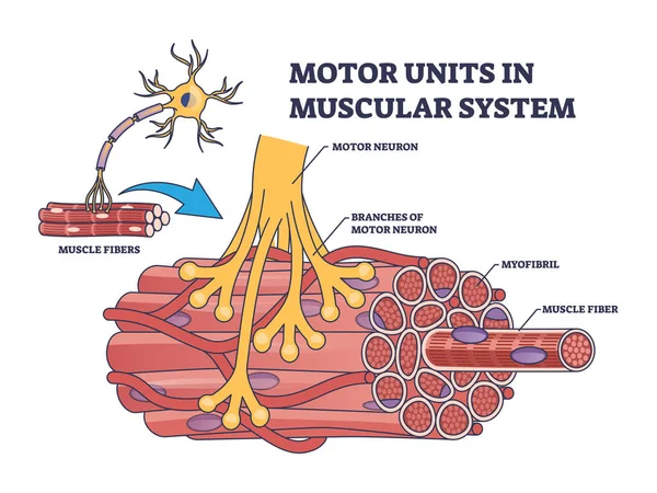
Motor Units In Muscular System With Fibers Neuron Anatomy Outline Diagram. Labeled Educational Medical Scheme With Myofibril And Muscle Fiber Closeup Vector Illustration. Nerve Functional Contraction
Vector, 6.89MB, 5000 × 3750 eps

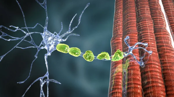
Motor Neuron Connecting To Muscle Fiber, 3D Illustration. A Neuromuscular Junction Allows The Motor Neuron To Transmit A Signal To The Muscle Causing Contraction. It Is Affected By Toxins And Diseases
Image, 11.3MB, 7200 × 4050 jpg
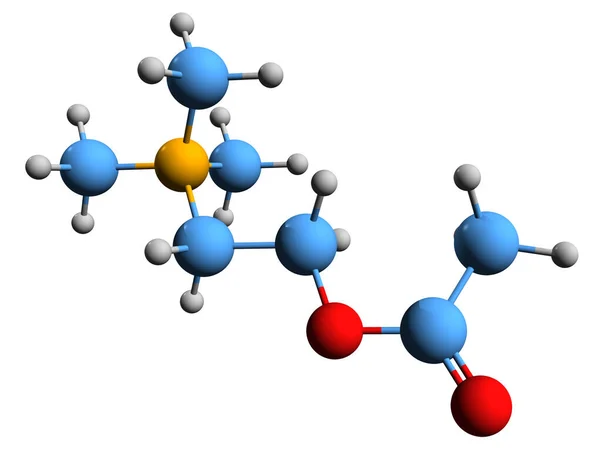
3D Image Of Acetylcholine Skeletal Formula - Molecular Chemical Structure Of Neurotransmitter ACh Isolated On White Background
Image, 1.87MB, 5000 × 3866 jpg
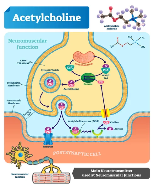
Acetylcholine Vector Illustration. Labeled Scheme With Neurotransmitter.
Vector, 11.74MB, 3700 × 4523 eps

Neuromuscular Spindle And Proprioception: Perception Of The Position Of Parts Of The Body.
Image, 0.76MB, 3630 × 3138 jpg
Neuromuscular Spindle And Proprioception: Perception Of The Position Of Parts Of The Body.
Image, 1.26MB, 4961 × 3543 jpg
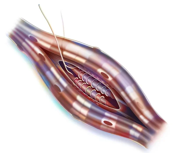
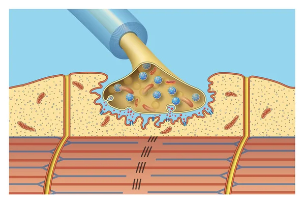
Neuromuscular Junction. A Synaptic Connection Between The Terminal End Of A Motor Nerve And A Muscle. Presynaptic (nerve Terminal), Postsynaptic Part, Synaptic Cleft.
Vector, 5.77MB, 6250 × 6250 eps
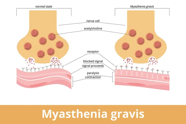
Myasthenia Gravis. An Autoimmune Disease Of The Neuromuscular Junction When Antibodies Block Or Destroy Nicotinic Acetylcholine Receptors (AChR) At The Junction Between The Nerve And Muscle.
Vector, 7.31MB, 6250 × 4167 eps

Human Anatomy Of A Motor Neuron, Including Its Parts Like The Axon And Dendrites Structure Diagram Hand Drawn Schematic Vector Illustration. Medical Science Educational Illustration
Vector, 0.6MB, 5000 × 3750 eps

Human Anatomy Of A Motor Neuron, Including Its Parts Like The Axon And Dendrites Structure Diagram Hand Drawn Schematic Raster Illustration. Medical Science Educational Illustration
Image, 3.52MB, 6000 × 4500 jpg

Detailed Structure Of Neuromuscular Junction, Vector Illustration For Neuroscience, Biology, And Medical Education On White Background
Vector, 0.45MB, 5000 × 3000 ai

Demyelination Of Neuron, The Damage Of The Neuron Myelin Sheath Seen In Demyelinating Diseases, 3D Illustration. Multiple Sclerosis And Other Demyelinating Myelinoclastic And Leukodystrophic Diseases
Image, 12.43MB, 7200 × 4050 jpg
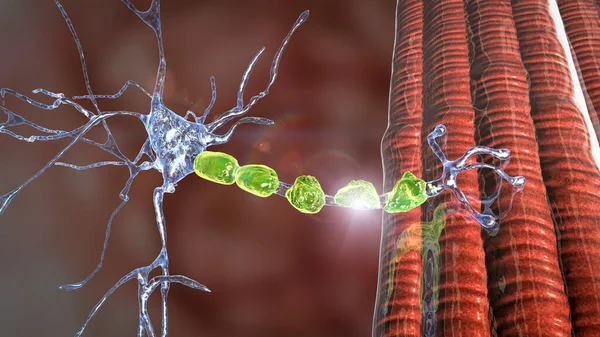
Motor Neuron Connecting To Muscle Fiber, 3D Illustration. A Neuromuscular Junction Allows The Motor Neuron To Transmit A Signal To The Muscle Causing Contraction. It Is Affected By Toxins And Diseases
Image, 10.6MB, 7200 × 4050 jpg
Demyelination Of Neuron, The Damage Of The Neuron Myelin Sheath Seen In Demyelinating Diseases, 3D Illustration. Multiple Sclerosis And Other Demyelinating Myelinoclastic And Leukodystrophic Diseases
Image, 12.46MB, 7200 × 4050 jpg
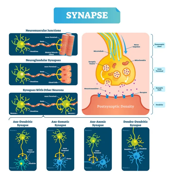
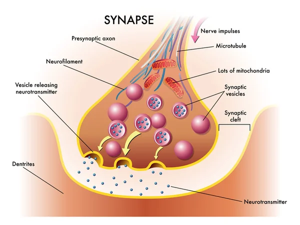
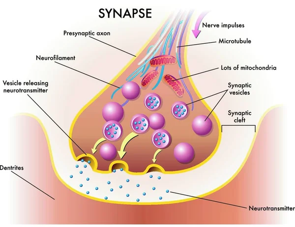
Synapse Vector Illustration. Labeled Diagram With Neuromuscular Example.
Vector, 7.34MB, 4000 × 4109 eps
Myasthenia Gravis Icon. Trendy Flat Vector Myasthenia Gravis Icon On White Background From Diseases Collection, Vector Illustration Can Be Use For Web And Mobile, Eps10
Vector, 0.56MB, 6944 × 6944 eps
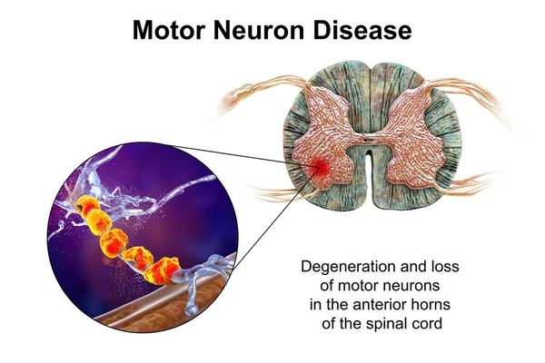
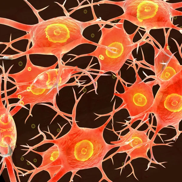
Motor Neuron Diseases, 3D Illustration Showing Degeneration Of Motor Neurons In Anterior Horns Of Spinal Cord. Amyotrophic Lateral Sclerosis And Other Motor Neuron Disorders
Image, 16.37MB, 10308 × 6872 jpg

Motor Neuron Diseases, 3D Illustration Showing Degeneration Of Motor Neurons In Anterior Horns Of Spinal Cord. Amyotrophic Lateral Sclerosis And Other Motor Neuron Disorders
Image, 14.98MB, 8408 × 5605 jpg
Demyelination Of Neuron, The Damage Of The Neuron Myelin Sheath Seen In Demyelinating Diseases, 3D Illustration. Multiple Sclerosis And Other Demyelinating Myelinoclastic And Leukodystrophic Diseases
Image, 17.84MB, 7200 × 4050 jpg

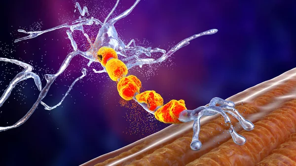
Motor Neuron Connecting To Muscle Fiber, 3D Illustration. A Neuromuscular Junction Allows The Motor Neuron To Transmit A Signal To The Muscle Causing Contraction. It Is Affected By Toxins And Diseases
Image, 16.17MB, 7200 × 4050 jpg
Myasthenia Gravis Icon. Trendy Modern Flat Linear Vector Myasthenia Gravis Icon On White Background From Thin Line Diseases Collection, Editable Outline Stroke Vector Illustration
Vector, 0.6MB, 6944 × 6944 eps

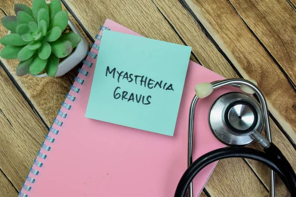
Concept Of Myasthenia Gravis Write On Sticky Notes Isolated On Wooden Table.
Image, 4.52MB, 6000 × 4000 jpg

Motor Neuron Diseases, 3D Illustration Showing Degeneration Of Motor Neurons In Anterior Horns Of Spinal Cord. Amyotrophic Lateral Sclerosis And Other Motor Neuron Disorders
Image, 12.99MB, 10308 × 6872 jpg

Demyelination Of Neuron, The Damage Of The Neuron Myelin Sheath Seen In Demyelinating Diseases, 3D Illustration. Multiple Sclerosis And Other Demyelinating Myelinoclastic And Leukodystrophic Diseases
Image, 24.2MB, 7200 × 4050 jpg


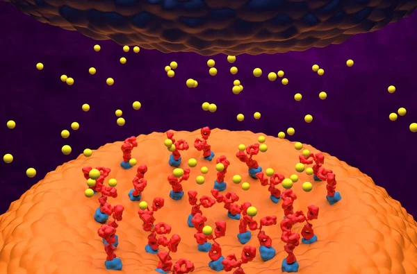
Autoantibodies Bond To Receptor (achr) Blocking The Acetylcholine Transmitters In Myasthenia Gravis (MG) - 3d Illustration Isometric View
Image, 6.74MB, 10000 × 6600 jpg
Motor Neuron Connecting To Muscle Fiber, 3D Illustration. A Neuromuscular Junction Allows The Motor Neuron To Transmit A Signal To The Muscle Causing Contraction. It Is Affected By Toxins And Diseases
Image, 18.96MB, 7200 × 4050 jpg
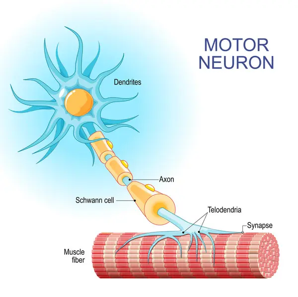
Motor Neuron. Structure And Anatomy Of A Efferent Neuron. Close-up Of A Muscle Fiber, And Motoneuron With Dendrites, Synapse, Telodendria, Axon, Schwann Cell. The Axons Carry Signals From The Spinal Cord To Muscles. Vector Illustration
Vector, 5.05MB, 4444 × 4422 eps

Demyelination Of Neuron, The Damage Of The Neuron Myelin Sheath Seen In Demyelinating Diseases, 3D Illustration. Multiple Sclerosis And Other Demyelinating Myelinoclastic And Leukodystrophic Diseases
Image, 20.55MB, 7200 × 4050 jpg

Myasthenia Gravis. Autoimmune Disease. Space Between Neuron And Muscle. In Myasthenia Gravis, Abnormal Antibodies Prevent Acetylcholine From Binding, Blocked Ion Channels And The Normal Communication Process Between Muscles And Nerves. Vector Poster.
Vector, 0.93MB, 5000 × 3965 eps
Autoantibodies Bond To Receptor (achr) Blocking The Acetylcholine Transmitters In Myasthenia Gravis (MG) - 3d Illustration Closeup View
Image, 14.17MB, 10000 × 6600 jpg
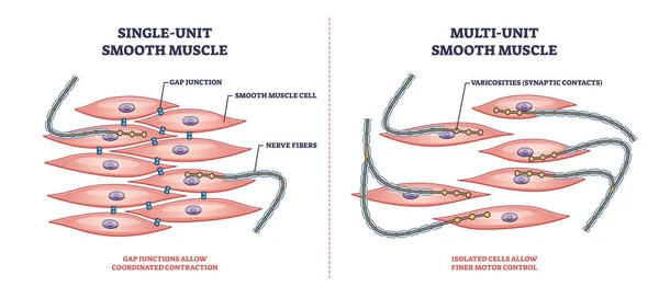
Single Unit Vs Multi Unit Smooth Muscle Structure Differences Outline Diagram
Vector, 6.26MB, 6000 × 2727 eps
Degradation Of Motor Neurons, Conceptual 3D Illustration. Motor Neuron Diseases Are A Group Of Neurodegenerative Disorders Including Amyotrophic Lateral Sclerosis, Progressive Bulbar Palsy And Other
Image, 10.87MB, 7200 × 4050 jpg

Motor Neuron Connecting To Muscle Fiber, 3D Illustration. A Neuromuscular Junction Allows The Motor Neuron To Transmit A Signal To The Muscle Causing Contraction. It Is Affected By Toxins And Diseases
Image, 15.53MB, 6075 × 4050 jpg

Page 1 >> Next