photo free
Home Images Vectors Videos

Stock image Ocular Histology

stock image Deep Dive: Intense View of Retina Layers at 400x

Deep Dive: Intense View Of Retina Layers At 400x

Image, 17.1MB, 4683 × 3512 jpg
stock image Periphery Revealed: Detailed Peripheral Retina Layers at 400x

Periphery Revealed: Detailed Peripheral Retina Layers At 400x

Image, 13.99MB, 4591 × 3443 jpg
stock image The daily life of a child with disabilities. A boy with Down syndrome plays with dogs. Chromosomal and genetic disorder in the baby.

The Daily Life Of A Child With Disabilities. A Boy With Down Syndrome Plays With Dogs. Chromosomal And Genetic Disorder In The Baby.

Image, 6.59MB, 4368 × 2912 jpg
stock image The daily life of a child with disabilities. A boy with Down syndrome plays with dogs. Chromosomal and genetic disorder in the baby.

The Daily Life Of A Child With Disabilities. A Boy With Down Syndrome Plays With Dogs. Chromosomal And Genetic Disorder In The Baby.

Image, 5.57MB, 4368 × 2912 jpg
stock image Retina layers, light micrograph. From top to bottom: pigment epithelium, rods and cones, outer nuclear, outer plexiform, inner nuclear, inner plexiform, ganglion cell, and nerve fibre layers. A dilated artery and a vein appear in the ganglion cell la

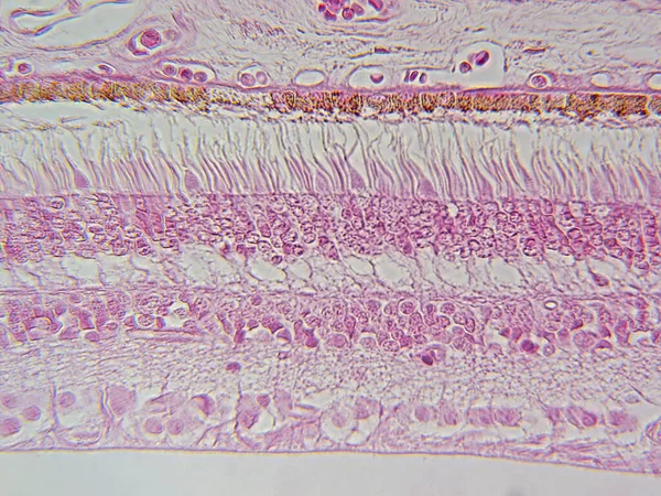
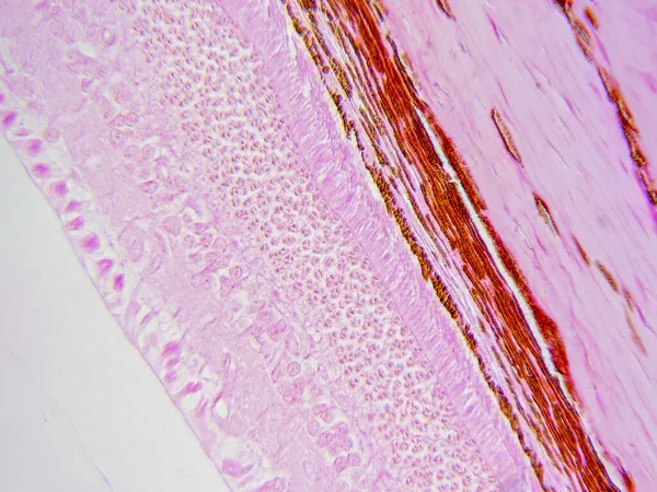
Retina Layers, Light Micrograph. From Top To Bottom: Pigment Epithelium, Rods And Cones, Outer Nuclear, Outer Plexiform, Inner Nuclear, Inner Plexiform, Ganglion Cell, And Nerve Fibre Layers. A Dilated Artery And A Vein Appear In The Ganglion Cell La

Image, 9.2MB, 3840 × 3072 jpg
Page 1 >> Next