Home
Images
Vectors
Videos
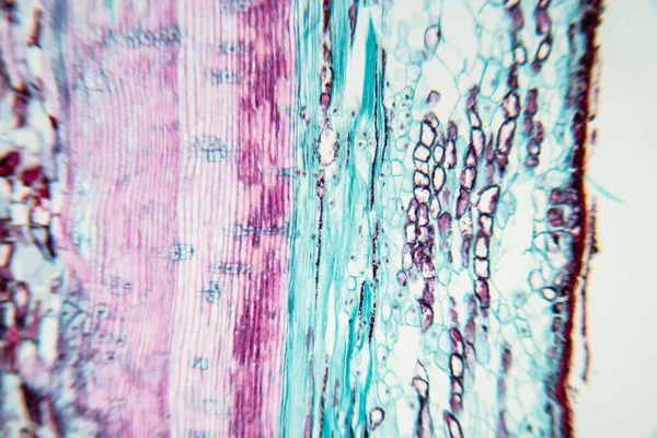
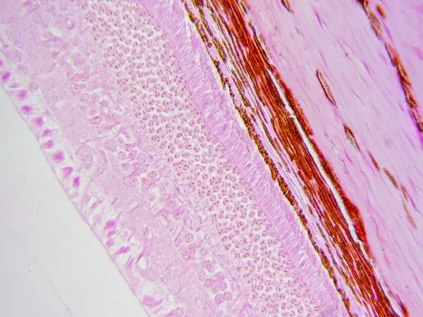
Stock image Periphery Revealed: Detailed Peripheral Retina Layers at 400x
Published:
May.23, 2023 14:12:53
Author:
DrWD40
Views:
3
Downloads:
1
File type:
image
/
jpg
File size:
13.99 MB
Orginal size:
4591
x
3443
px
Available sizes:
S
(500 x 375)
M
(1000 x 750)
L
(2000 x 1500)
XL
(4591 x 3443)
EL0
(4591 x 3443)
Level:
beginner
Download Image
Image topics:
Education
Biology
Ophthalmology
Histology
Medical Research
Eye Health
Cellular Structure
Histopathology
Eye Anatomy
Neuro Ophthalmology
Retinal Cells
Biomedical Imaging
400x Magnification
Retinal Tissue
Photoreceptor Cells
Anatomical Science
Histological Sample
Ocular Anatomy
Ocular Histology
Retinal Layers
Vision Biology
Vision Science
Peripheral Retina
Peripheral Retina Layers
Similar stock images
Plant Vascular Tissue Under The Microscope View For Education.
6000 × 4000
The Study Plant Tissue Of Under The Microscope For Classroom Education.
6720 × 4480
Petals Of A Tulip Close-up Background Texture
4580 × 3034
The Study Plant Tissue Of Under The Microscope For Classroom Education.
6720 × 4480
Histology Of Human Compact Bone Tissue Under Microscope View For Education, Muscle Bone Connection And Connective Tissue
6000 × 4000
Crayon Hand Drawing Purple Pink Texture For Background
5000 × 3333
Characteristics Of Fungi Living In Wood As A Group, Are Polyphyletic Under The Microscope For Education.
6720 × 4480
Purple Dirty Art Painting. Artistic Dirty Art. Wet Art Print. Aquarelle Texture. Splash Banner. Pink Watercolor Print. Indigo Tie Dye Print. Brushed Graffiti. Tie Dye Batik. Blue
5000 × 3183