
Stock image Optic Nerve page 3
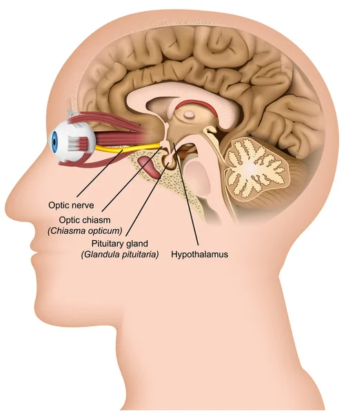
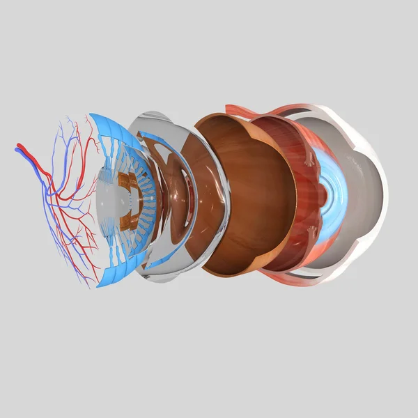
Optic Nerve Anatomy 3d Medical Vector Illustration On White Background
Vector, 19.02MB, 5000 × 6000 eps

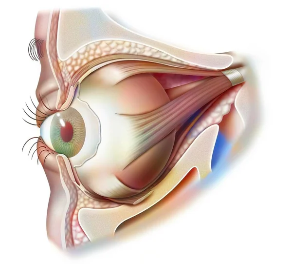
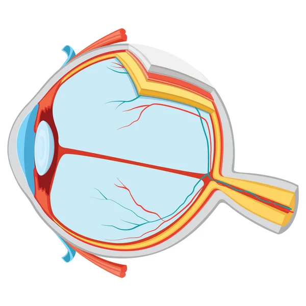
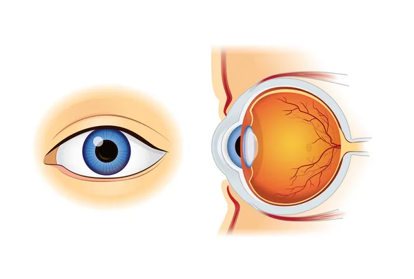
Eyes Are The Organs Of Vision. They Detect Light And Convert It Into Electro-chemical Impulses In Neurons. Illustration Anatomy Body.
Vector, 0MB, 3680 × 4144 zip

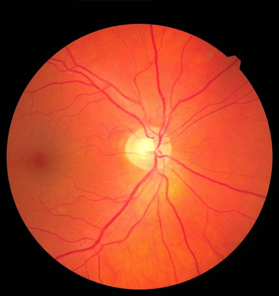
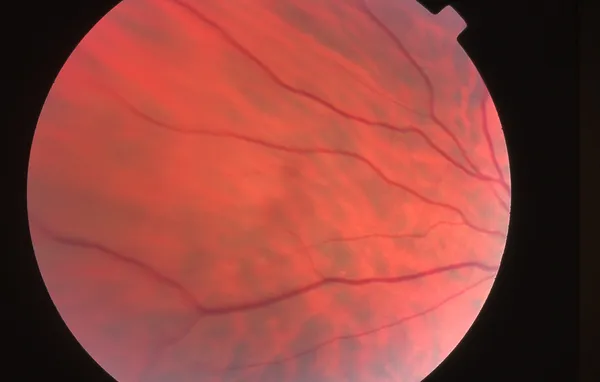
View Inside Human Eye Showing Retina, Optic Nerve And Macula. Health Concept
Image, 2.58MB, 3000 × 3186 jpg
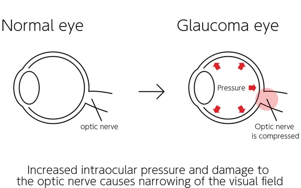
Illustration Of How Glaucoma Works, Normal And Glaucomatous Eyes, Medical Illustration
Vector, 0.1MB, 5334 × 3334 ai
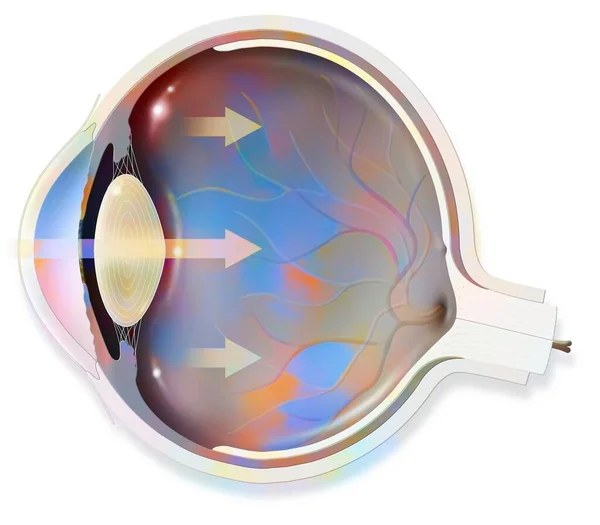
Anatomy Of The Eye Whose Arrows Represent Light And Revealing The Lens, Retina, Cornea, Iris, Choroid. .
Image, 0.76MB, 3630 × 3175 jpg
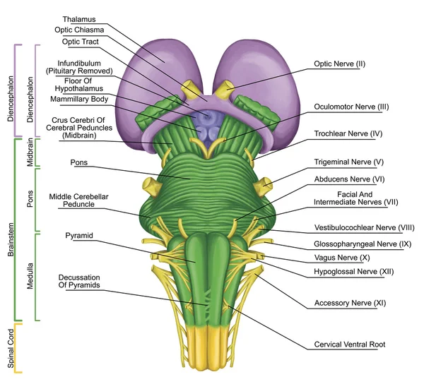
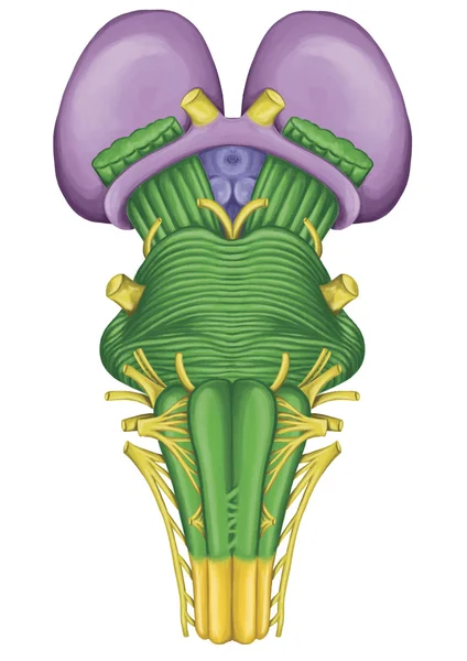
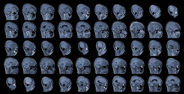
Brainstem, Brain Stem, Ventral View, Posterior Part Of The Brain, Adjoining And Structurally Continuous With The Spinal Cord, Motor And Sensory Innervation To The Face And Neck Via Thecranial Nerves
Image, 7.81MB, 7018 × 6452 jpg


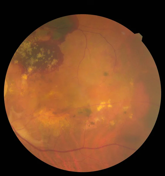
View Inside Human Eye Disorders Showing Retina, Optic Nerve And Macula. Retinal Picture ,Medical Photo Tractional Eye Screen Retinal Detachment Of Diabetes. Eye Treatment Concept.
Image, 3.37MB, 3000 × 3186 jpg
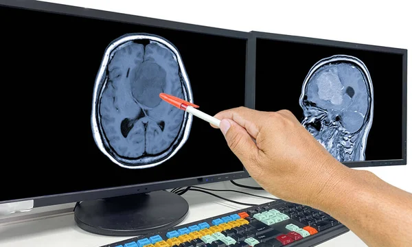
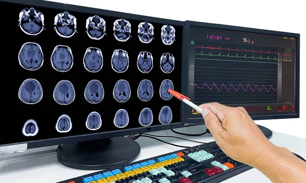
Glioblastoma, Brain Metastasis,MRI Brain The Doctor Pointed Out The Location Of The Brain Tumor On The Computer Screen
Image, 9.74MB, 7000 × 4205 jpg
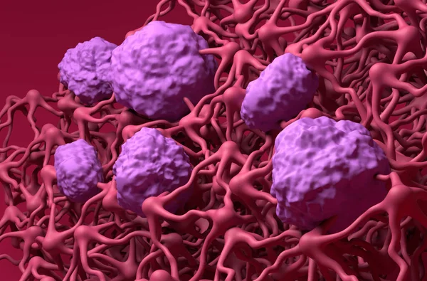
Blood-brain Barrier (BBB) In The Human Brain - Closeup View 3d Illustration
Image, 7.46MB, 10000 × 6600 jpg
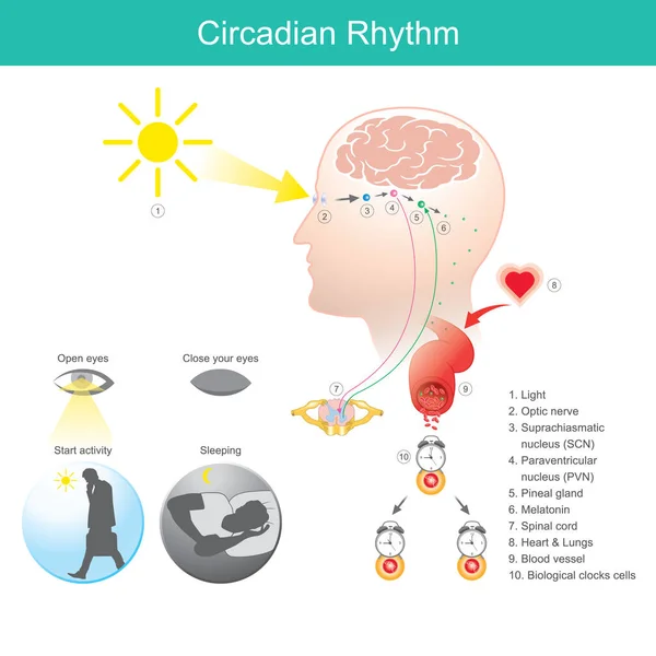
Circadian Rhythm. Diagram Human Body Physical, Mental, And Behavioural Changes That Follow A 24-hour Cycle
Vector, 10.55MB, 5000 × 5000 eps
Brainstem, Brain Stem, Ventral View, Adjoining And Structurally Continuous With The Spinal Cord, Parts Of The Diencephalon, Motor And Sensory Innervation To The Face And Neck Via Thecranial Nerves
Image, 3.55MB, 3508 × 4961 jpg

Ophthalmic Image Detailing The Retina And Optic Nerve Inside A Healthy Human Eye. Medicine Concept
Image, 3.76MB, 3000 × 3186 jpg

Eye Disease, Atrophic Age-Related Macular Degeneration Illustrated - Translation: Normal State, Atrophic Age-related Macular Degeneration, Atrophy Of Macular Tissue With Age, Retina, Choroid, Central Fossa (macula)
Vector, 0.25MB, 5334 × 3334 ai
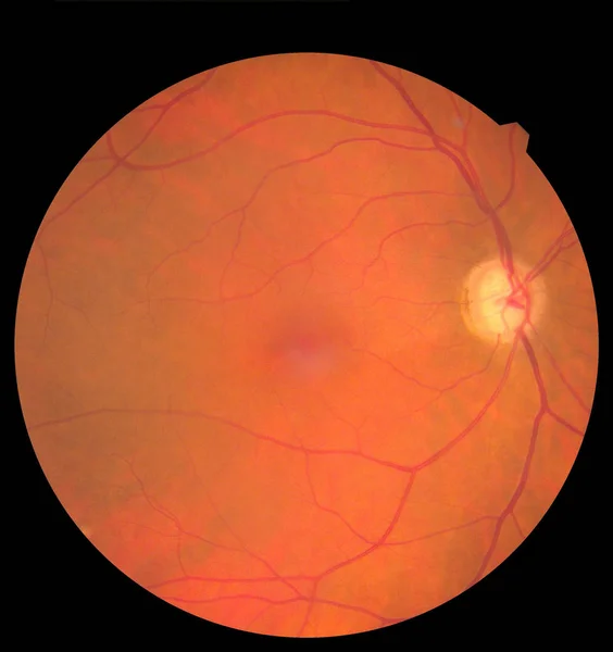
View Inside Human Eye Showing Retina, Optic Nerve And Macula. Health Concept
Image, 2.84MB, 3000 × 3186 jpg
MRI Brain Axial Views .to Evaluate Brain Tumor. Glioblastoma, Brain Metastasis Isodensity Mass With An Ill-defined Margin And Surrounding Edema At The Right Frontal Lobe.
Image, 10.68MB, 8183 × 4192 jpg

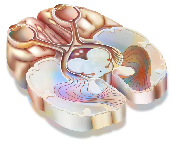
The Optic Tract: Transmission Of Visual Information From The Retina To The Visual Cortex.
Image, 0.97MB, 3630 × 3076 jpg

Illustration Of How Glaucoma Works, Glaucoma Eye, Medical Illustration
Vector, 0.06MB, 5334 × 3334 ai
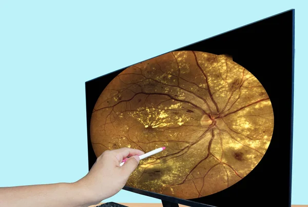
Close Up Hand Doctor Point LCD Monitor Report Screen Fundus Photography Of Diabetes.Medical Healthcare Concept
Image, 8.46MB, 6136 × 4140 jpg


Eyeball With Optic Nerve. Watercolor Illustration. Green Iris Realistic Eye Human Element. Close Up Eye Ball With Ocular Nerve. Human Face Round 3d Detail. Face Anatomy Element On White Background
Image, 3.96MB, 7000 × 4500 jpg

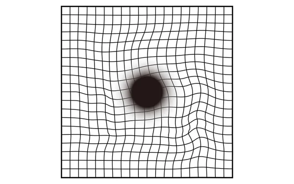
Amsler Chart, Age-related Macular Degeneration Visibility (visual Field Defects, Central Darkening)
Vector, 0.11MB, 5334 × 3334 ai
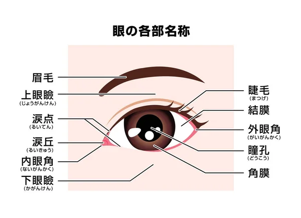
Structure Of Human Eye ( Names Of Parts ) Vector Illustration / Japanese
Vector, 5.82MB, 8055 × 5846 eps

The Patient Is A Doctor With An Ophthalmologist Who Is Watching An Eye Examination Using Modern Technology. Fundus And Optic Nerve Research Concept, Ophthalmoscopy
Image, 4.26MB, 5474 × 3396 jpg
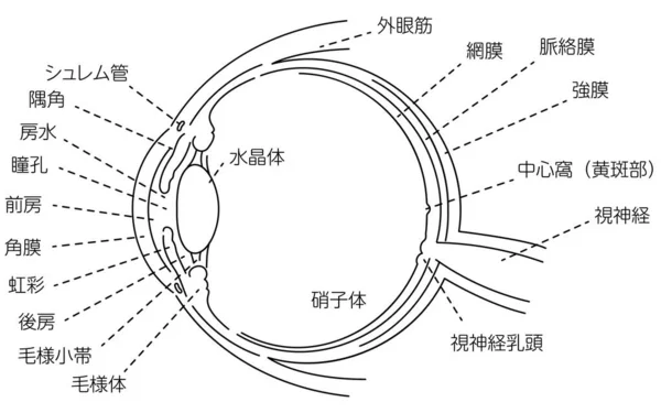
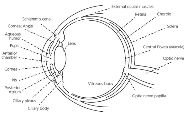
Illustration Of The Eye - Translation: Schlemm's Canal, Corner Angle, Aqueous Humor, Pupil, Anterior Chamber, Cornea, Iris, Posterior Chamber, Ciliary Body, Ciliary Body, Lens, Vitreous Body, External Eye Muscle, Retina, Choroid, Sclera, Central Foss
Vector, 0.19MB, 5334 × 3334 ai
View Inside Human Eye Disorders Showing Retina, Optic Nerve And Macula. Retinal Picture ,Medical Photo Tractional Eye Screen Retinal Detachment Of Diabetes. Eye Treatment Concept.
Image, 2.62MB, 3000 × 3186 jpg
Glioblastoma, Brain Metastasis,MRI Brain The Doctor Pointed Out The Location Of The Brain Tumor On The Computer Screen
Image, 4.5MB, 7000 × 4205 jpg


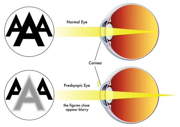
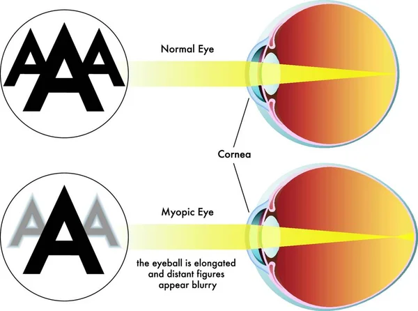
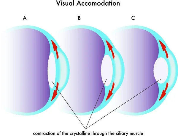
Illustration Of The Three Visual Defects Myopia, Hyperopia And Astigmatism And How To Correct It With Biconcave And Biconvex Lenses - With Glasses Or Contact Lenses.
Vector, 0MB, 7088 × 3396 zip


Illustration Of How Glaucoma Works, Normal And Glaucomatous Eyes, Medical Illustration - Translation: Normal Eye, Glaucoma Eye, Pressure, Optic Nerve, Optic Nerve Is Compressed, High Intraocular Pressure And Damage To Optic Nerve Causes Visual Field
Vector, 0.12MB, 5334 × 3334 ai
Previous << Page 3 >> Next