
Stock image Patellar Ligament


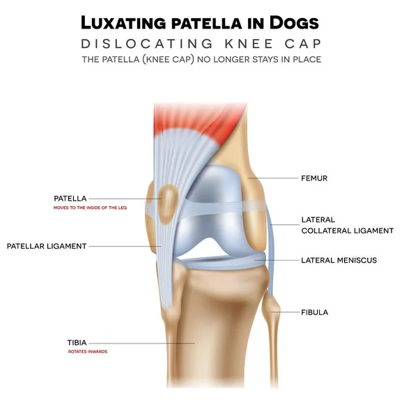
Luxating Patella In Dogs, It Shifts Either Towards The Inner Or Outer Knee. Anatomy Of The Canine (dog's) Knee Joint Colorful Design, Medical Info Poster Illustration.
Vector, 12.18MB, 5000 × 5000 eps

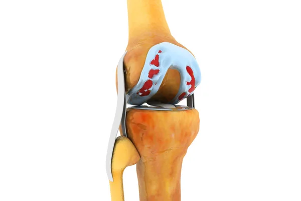
Unicompartmental Knee Arthroplasty. Surgical Procedure For Treatment Or Relieve Arthritis, After Joint Damaged. Uni Knee Implant. Partial Knee Replacement On Blue And White Background. Side View Of Human Joint. Vector Illustration
Vector, 4.48MB, 4444 × 4444 eps

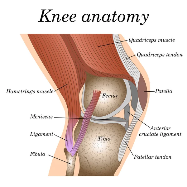
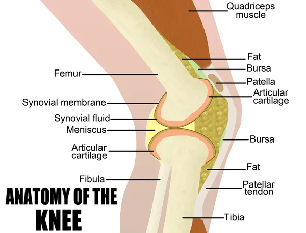
Knee Anatomy. Side And Front View. Cross Section Of The Joint Showing The Main Parts: Femur, Fibula, Articular Capsule, Menisci, Muscles And Ligaments. Vector Illustration
Vector, 3.91MB, 4444 × 3973 eps

Medial Knee Ligament Sprain Medical Vector Illustration Isolated On White Background Infographic
Vector, 7.79MB, 6000 × 5000 eps

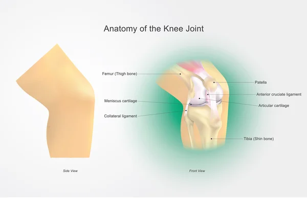
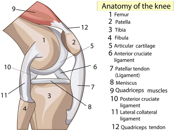
Anatomy Of The Knee Joint Front View, Template For Training A Medical Surgical Poster, Traumatology Page. Vector Illustration.
Vector, 5MB, 5000 × 5000 eps

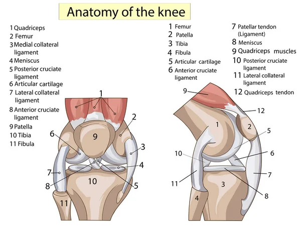
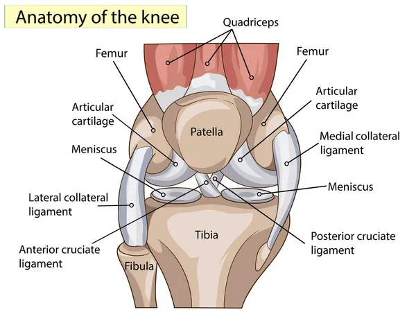
Knee Anatomy. Structure Of Leg Joint. Major Parts. Vector Poster With Text Label For Medical Education
Vector, 8.29MB, 4444 × 4444 eps

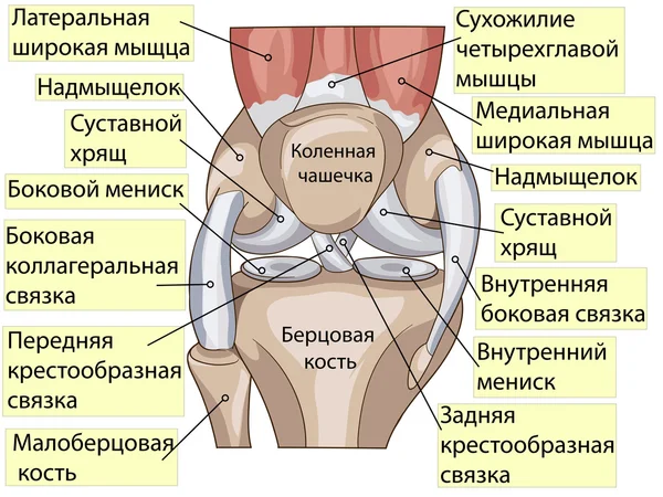
Structure Of The Human Knee Joint With The Name And Description Of All Sites. Lateral View. Medical Science Anatomy Poster. Vector Illustration Isolated On White Background.
Vector, 6.98MB, 8334 × 8334 eps
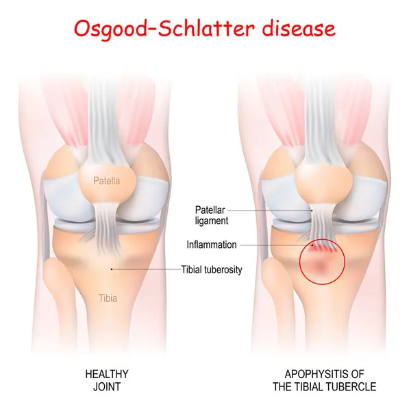
Osgood-Schlatter Disease. Healthy Joint And Apophysitis Of The Tibial Tubercle. Vector Illustration. Poster For Medical Use
Vector, 3.08MB, 4444 × 4445 eps

Anatomical Design. Posterior And Radial Collateral Ligament Of The Elbow Joint.
Image, 3.06MB, 6000 × 4500 jpg
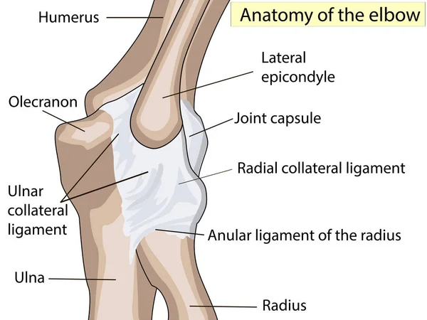
Anatomical Design. Posterior And Radial Collateral Ligament Of The Elbow Joint.
Vector, 0.59MB, 5333 × 4000 eps
Anatomy Of The Knee Joint Side View, Template For Training A Medical Surgical Poster, Traumatology Page. Vector Illustration.
Vector, 2.75MB, 5000 × 5000 eps

Fibular Collateral Ligament Injury. Joint Anatomy. Vector Illustration For Biological, Medical, Science And Educational Use
Vector, 8.67MB, 5470 × 5470 eps

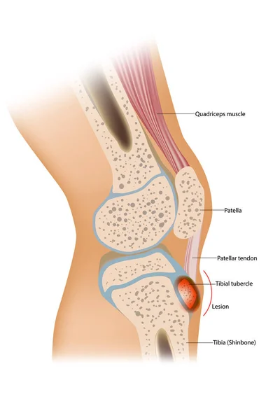
Osgood Schlatter Disease Or OSD Is Inflammation Of The Patellar Ligament At The Tibial Tuberosity
Vector, 4.44MB, 3900 × 5850 eps


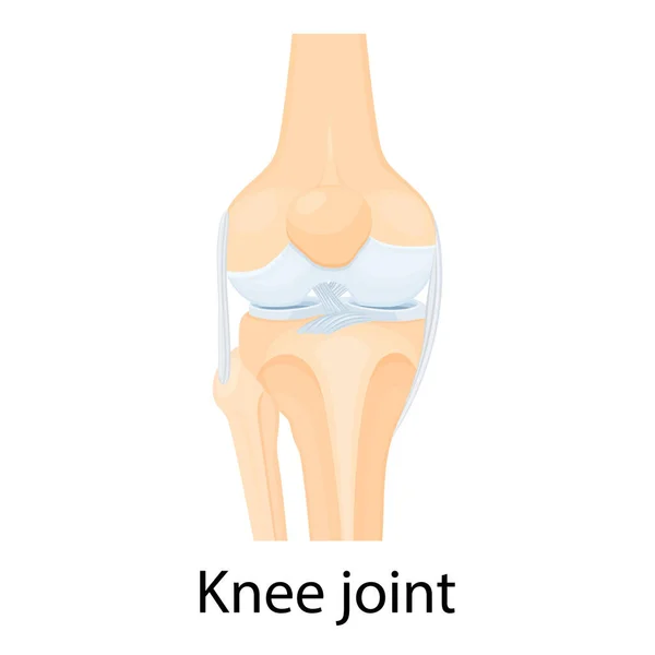
Knee Joint Isolated Vector Illustration, Flat Design. Ligaments Of The Knee. Anterior And Posterior Cruciate Ligaments, Patellar And Quadriceps, Tendons, Medial And Lateral Collateral Ligaments.
Vector, 1.01MB, 5000 × 5000 eps

Anatomy. Subscribe. Structure Knee Joint Raster Basic Medical Education
Image, 3.63MB, 6000 × 4500 jpg


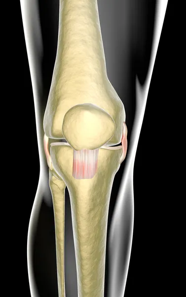
Knee Tendons Medical Vector Illustration Scheme, Anatomical Diagram.
Vector, 4.34MB, 4892 × 4004 eps
Anatomy. Subscribe. Structure Knee Joint Raster Basic Medical Education
Image, 3.47MB, 6000 × 4500 jpg


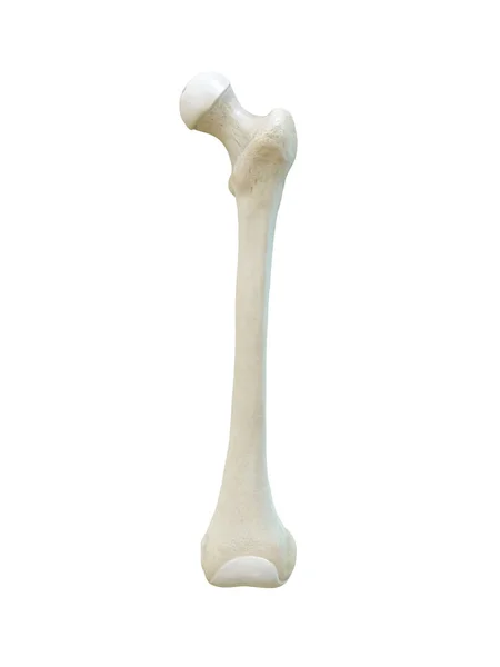
Left Human Femur Bone, Anterior View, Bone Anatomy, White Background, 3d Rendering
Image, 0.58MB, 3375 × 4500 jpg

Page 1 >> Next