photo free
Home Images Vectors Videos

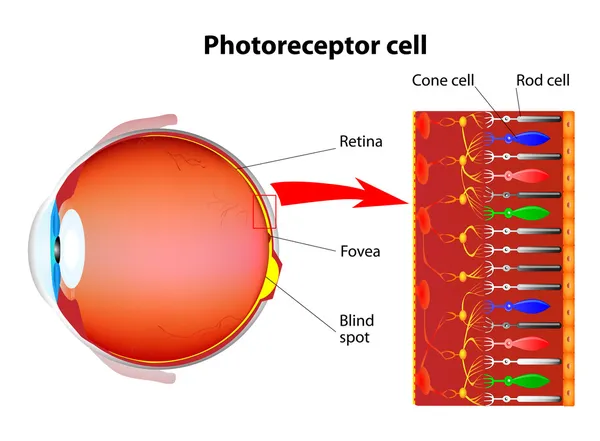
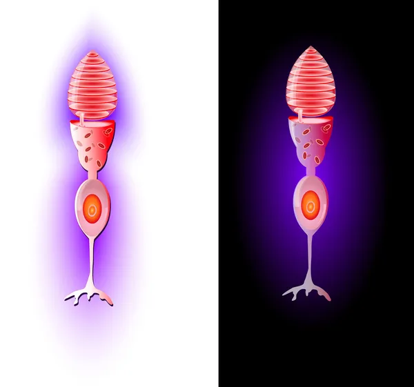
Stock image Photoreceptor Cells

stock vector Anatomy of Photoreceptor. cell of a retina in the eye. Cone cells in respond to color vision and send signals to brain. Rod cells are used in peripheral vision

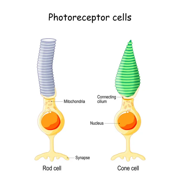
Anatomy Of Photoreceptor. Cell Of A Retina In The Eye. Cone Cells In Respond To Color Vision And Send Signals To Brain. Rod Cells Are Used In Peripheral Vision

Vector, 10.49MB, 4444 × 4444 eps
stock vector Structure of the human eye and organization of the retina. Optic part of retina.

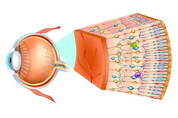
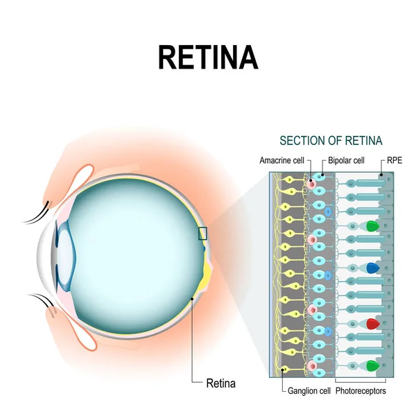
Structure Of The Human Eye And Organization Of The Retina. Optic Part Of Retina.

Vector, 8.08MB, 5001 × 3334 eps
stock image Deep Dive: Intense View of Retina Layers at 400x

Deep Dive: Intense View Of Retina Layers At 400x

Image, 17.1MB, 4683 × 3512 jpg
stock image Periphery Revealed: Detailed Peripheral Retina Layers at 400x

Periphery Revealed: Detailed Peripheral Retina Layers At 400x

Image, 13.99MB, 4591 × 3443 jpg
stock vector Biological diagram of rod and cone cells in retina

Biological Diagram Of Rod And Cone Cells In Retina

Vector, 42.97MB, 4273 × 3793 eps
stock vector Retina structure. Retina cell organization including rods and cones, horizontal cells, retinal pigment epithelium, Muller and ganglion cell. Retina histology (human eye).

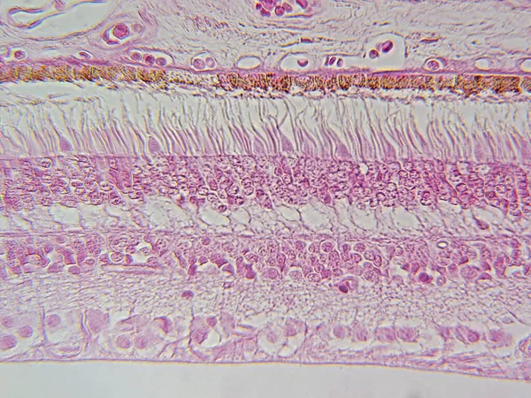
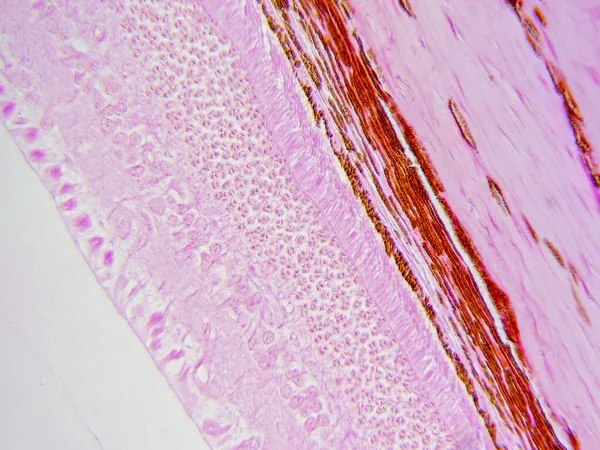
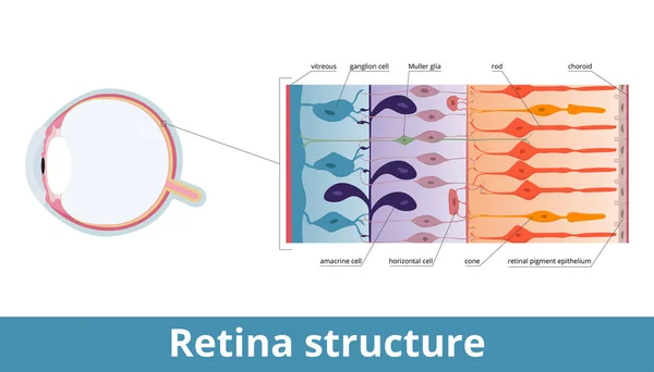
Retina Structure. Retina Cell Organization Including Rods And Cones, Horizontal Cells, Retinal Pigment Epithelium, Muller And Ganglion Cell. Retina Histology (human Eye).

Vector, 5.71MB, 7292 × 4167 eps
stock vector The Rod cell. Model Photoreceptor. Vector

The Rod Cell. Model Photoreceptor. Vector

Vector, 10.24MB, 4400 × 3629 eps
stock vector Retina. Rod cells and cone cells. Vector

Retina. Rod Cells And Cone Cells. Vector

Vector, 13.23MB, 4923 × 3807 eps
stock vector Photoreceptor cells in the retina of the eye

Photoreceptor Cells In The Retina Of The Eye

Vector, 1.83MB, 5473 × 4025 eps
stock vector retinal cells: rod and cone cells

Retinal Cells: Rod And Cone Cells

Vector, 3.07MB, 4594 × 4594 eps
stock vector Eye anatomy. Rod cells and cone cells. The arrangement of retinal cells is shown in a cross section. Vector diagram for your design, educational, biological, science and medical use

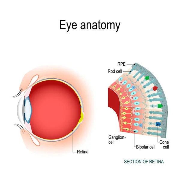
Eye Anatomy. Rod Cells And Cone Cells. The Arrangement Of Retinal Cells Is Shown In A Cross Section. Vector Diagram For Your Design, Educational, Biological, Science And Medical Use

Vector, 3.23MB, 5154 × 5154 eps
stock vector Cone cells. Vector

Cone Cells. Vector

Vector, 12.72MB, 5129 × 4798 eps
stock vector CPK - creatine phosphokinase acronym

CPK - Creatine Phosphokinase Acronym

Vector, 3.39MB, 5000 × 3750 eps
stock vector Melatonin and  Circadian rhythm regulation. Brain with pineal gland and suprachiasmatic nucleus. sleep-wake cycle. Human anatomy. vector illustration. What does melatonin do?

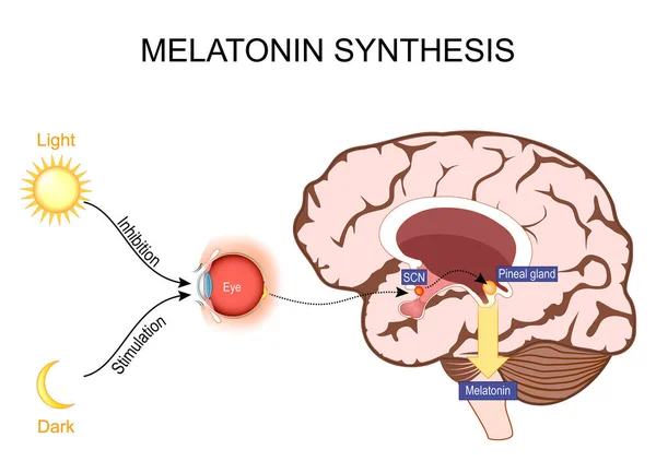
Melatonin And Circadian Rhythm Regulation. Brain With Pineal Gland And Suprachiasmatic Nucleus. Sleep-wake Cycle. Human Anatomy. Vector Illustration. What Does Melatonin Do?

Vector, 2.72MB, 5000 × 3613 eps
stock vector Rod Cell business card design

Rod Cell Business Card Design

Vector, 0.71MB, 6944 × 6944 eps
Page 1 >> Next