
Stock image Preretinal

Proliferative Diabetic Retinopathy, Illustration Showing Neovascularization In The Disk And Macula Edema. Abnormal Finding On Fundoscopic Examination Of The Eye Retina In Diabetes Mellitus
Image, 2.85MB, 5000 × 5000 jpg
Proliferative Diabetic Retinopathy, Illustration Showing Neovascularization In The Disk And Other Sites, And Macula Edema. Fundoscopic Examination Of The Eye Retina In Diabetes Mellitus
Image, 2.9MB, 5000 × 5000 jpg
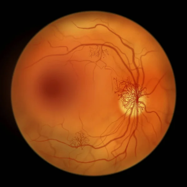
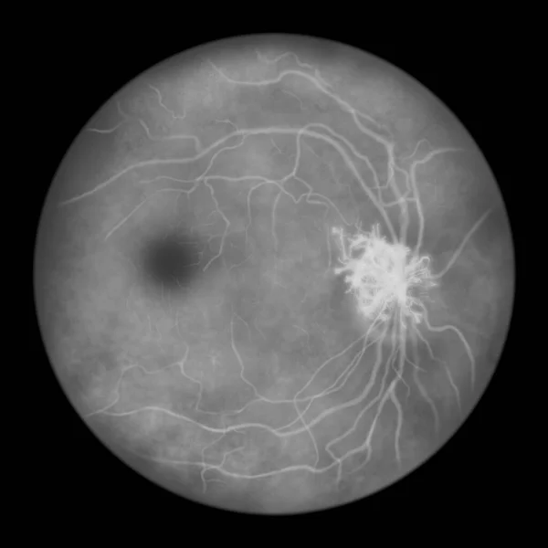
Proliferative Diabetic Retinopathy, Illustration Showing Neovascularization (formation Of New Vessels) In The Optic Disk. Fundoscopic Examination Of The Eye Retina In Diabetes Mellitus
Image, 2.81MB, 5000 × 5000 jpg

Proliferative Diabetic Retinopathy, Illustration Showing Neovascularization In The Optic Disk And Other Sites. Fundoscopic Examination Of The Eye Retina In Diabetes Mellitus, Fluorescein Angiography
Image, 6.07MB, 11738 × 6603 jpg

Proliferative Diabetic Retinopathy, Illustration Showing Neovascularization In The Disk And Other Sites, And Macula Edema. Eye Retina In Diabetes Mellitus, Fluorescein Angiography
Image, 2.67MB, 5000 × 5000 jpg
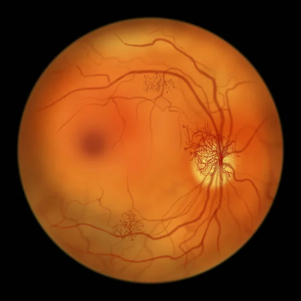
Proliferative Diabetic Retinopathy, Illustration Showing Neovascularization (formation Of New Vessels) In The Optic Disk And Other Sites. Fundoscopic Examination Of The Eye Retina In Diabetes Mellitus
Image, 2.86MB, 5000 × 5000 jpg

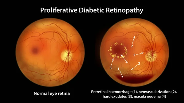
Proliferative Diabetic Retinopathy, 3D Illustration Showing Preretinal Haemorrhage As Horizontal Blood Level, Neovascularization In The Disk And Other Sites, Macula Edema And Hard Exudates
Image, 22.98MB, 11738 × 6603 jpg
Proliferative Diabetic Retinopathy, Illustration Showing Preretinal Haemorrhage As Horizontal Blood Level, Neovascularization In The Disk And Other Sites, Macula Edema And Hard Exudates
Image, 7.07MB, 11738 × 6603 jpg
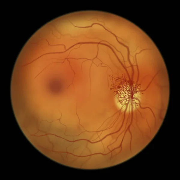
Proliferative Diabetic Retinopathy, Illustration Showing Neovascularization (formation Of New Vessels) In The Optic Disk. Eye Retina In Diabetes Mellitus, Fluorescein Angiography
Image, 2.63MB, 5000 × 5000 jpg
Page 1 >> Next