Home
Images
Vectors
Videos
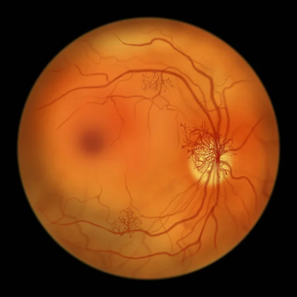
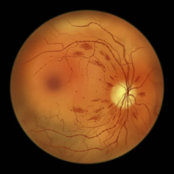
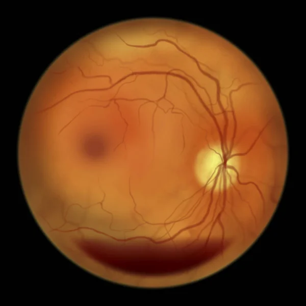
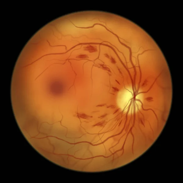
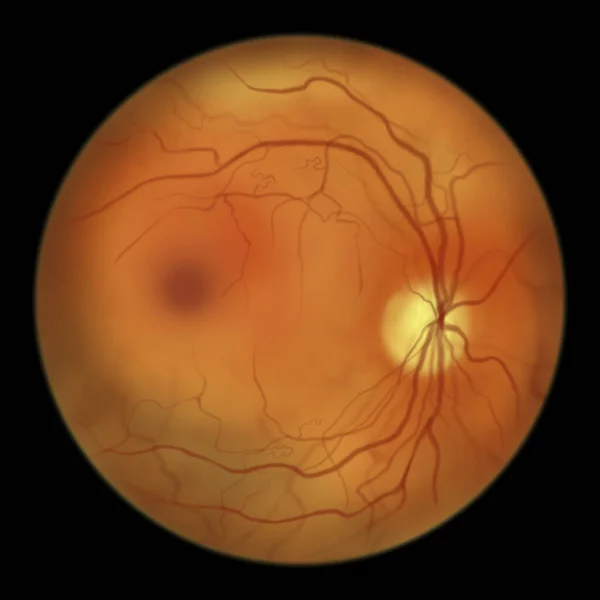
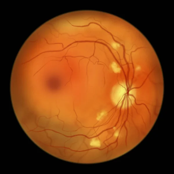
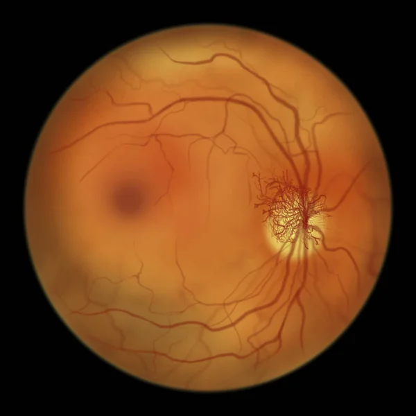
Stock image Proliferative diabetic retinopathy, illustration showing neovascularization (formation of new vessels) in the optic disk. Fundoscopic examination of the eye retina in diabetes mellitus
Published:
May.16, 2022 07:28:20
Author:
katerynakon
Views:
21
Downloads:
1
File type:
image
/
jpg
File size:
2.81 MB
Orginal size:
5000
x
5000
px
Available sizes:
S
(500 x 500)
M
(1000 x 1000)
L
(2000 x 2000)
XL
(5000 x 5000)
EL0
(5000 x 5000)
Level:
silver
Download Image
Image topics:
Illustration
Growth
Form
Medical
Eye
Clinical
Exam
Disease
Damaged
Disk
Diagnosis
Diagnostic
Ophthalmic
Ocular
Ophthalmology
Eyeball
Diabetic
Capillary
Bleeding
Diabetes
Retina
Ophthalmoscopy
Retinal
Capillaries
Ophthalmoscope
Fundus
Macula
Retinopathy
Hand Drawn
Hand Drawing
Blood Vessels
Proliferative
Blood Vessel
Diabetic Retinopathy
Diabetes Mellitus
Optic Disc
Eye Disease
Retinoscopy
Neovascularization
Fundoscopy
Optic Disk
Fundus Camera
Fundoscopic
Intraretinal
Preretinal
Similar stock images
Proliferative Diabetic Retinopathy, Illustration Showing Neovascularization (formation Of New Vessels) In The Optic Disk And Other Sites. Fundoscopic Examination Of The Eye Retina In Diabetes Mellitus
5000 × 5000
Non-proliferative Diabetic Retinopathy, Illustration Showing IRMAs (intraretinal Microvascular Abnormalities), Microaneurysms, Dot Haemorrhages, Flame-shaped And Splinter Retinal Haemorrhages
5000 × 5000
Non-proliferative Diabetic Retinopathy, Illustration Showing Microaneurysms, Dot Haemorrhages, Flame-shaped And Splinter Retinal Haemorrhages, Ophthalmoscope View
5000 × 5000
Diabetic Retinopathy, Illustration Shows Preretinal Haemorrhage As Horizontal Blood Level (boat-shaped Haemorrhage), Abnormal Finding On Fundoscopic Examination Of The Eye Retina In Diabetes Mellitus
5000 × 5000
Non-proliferative Diabetic Retinopathy, Illustration Showing Flame-shaped And Splinter Retinal Haemorrhages, Ophthalmoscope View
5000 × 5000
Non-proliferative Diabetic Retinopathy, Illustration Showing IRMAs (intraretinal Microvascular Abnormalities) As Small Vessels With Abnormal Branching Or Dilatation In Ischaemic Areas
5000 × 5000
Non-proliferative Diabetic Retinopathy, Illustration Showing Cotton Wool Spots As Fluffy Yellow Patches, Abnormal Finding On Funduscopic Examination Of The Eye Retina In Diabetes Mellitus
5000 × 5000
Proliferative Diabetic Retinopathy, Illustration Showing Neovascularization In The Disk And Macula Edema. Abnormal Finding On Fundoscopic Examination Of The Eye Retina In Diabetes Mellitus
5000 × 5000