
Stock image Pulmonary Embolism Diagnosis


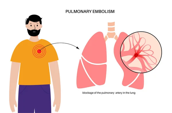
Pulmonary Embolism Disease. Deep Vein Thrombosis In Male Cartoon Body. Blood Clot In Lung Vein Anatomical Poster. Blocking Blood Flow In Lungs. Circulatory System Problem. DVT Vector Illustration
Vector, 0.48MB, 5883 × 3865 eps
Pulmonary Embolism Disease. Deep Vein Thrombosis. Blood Clot In Lung Vein Close Up Anatomical Poster. Blocking Blood Flow In Lungs. Circulatory System Problem. DVT Medical Flat Vector Illustration
Vector, 0.51MB, 4584 × 3789 eps

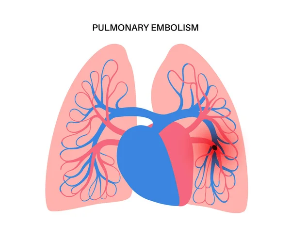
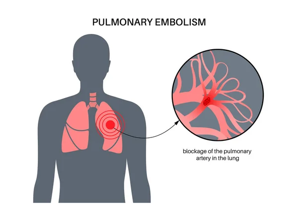
Pulmonary Embolism Vector Illustration. Labeled Lung Blood Blockage Scheme
Vector, 11.34MB, 4000 × 4000 eps


Base Of Lung 3D Rendering Image For Diagnosis TB,tuberculosis And Covid-19 From CT-Scanner 3D.
Image, 3.73MB, 5576 × 4056 jpg
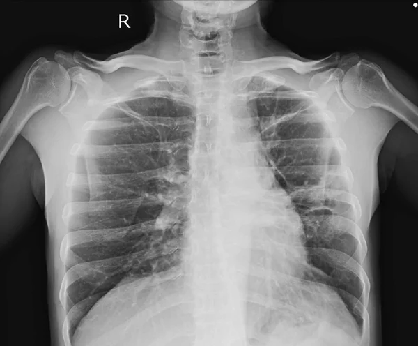
Chest X-ray Showing A 3 Cm Pleural Based Nodule At Lt Upper Lobe With Splicated Margin. Thickening Of Nearby Pleural. Nodular Infiltration At RUL. Normal Heart Size. Differential Diagnosis CA Lung, TB Granuloma.
Image, 2.36MB, 3637 × 3152 jpg
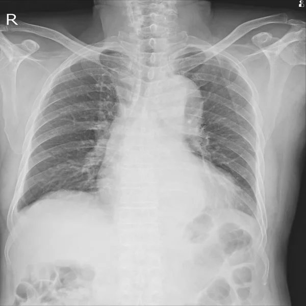
X-ray Chest Showing.Severe Cardiomegaly. Both Costophrenic Angles Are Clear.Intact Osseous StructureSevere Cardiomegaly.Infiltration At Lt Lower DDx. Pneumonia, Pulmonary Edema
Image, 3.88MB, 3800 × 3800 jpg
Chest X-ray Showing A 3 Cm Pleural Based Nodule At Lt Upper Lobe With Splicated Margin. Thickening Of Nearby Pleural. Nodular Infiltration At RUL. Normal Heart Size. Differential Diagnosis CA Lung, TB Granuloma.
Image, 2.09MB, 3628 × 3000 jpg


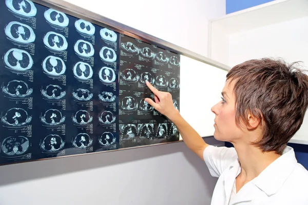
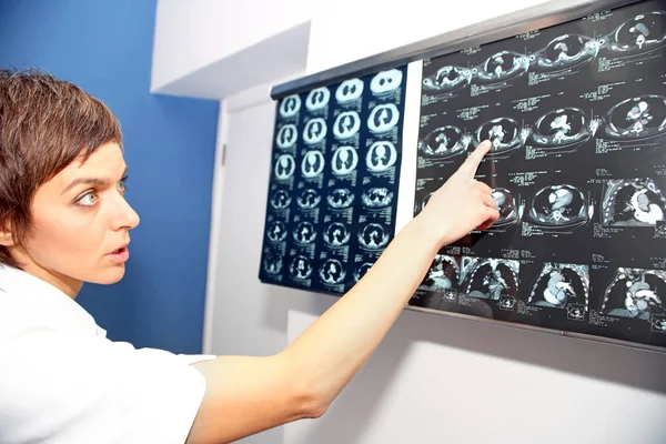
Doctor At Appointment Inspects And Examines Ct Scan Of Chest And Abdominal Cavity. Concept Photo On Diagnosis Of Diseases In Pulmonology, Lung Pathology, Inflammatory Diseases Of Bronchi, Tuberculosis
Image, 5.88MB, 3648 × 5472 jpg

Doctor At Appointment Inspects And Examines Ct Scan Of Chest And Abdominal Cavity. Concept Photo On Diagnosis Of Diseases In Pulmonology, Lung Pathology, Inflammatory Diseases Of Bronchi, Tuberculosis
Image, 6.94MB, 3648 × 5472 jpg

Doctor At Appointment Inspects And Examines Ct Scan Of Chest And Abdominal Cavity. Concept Photo On Diagnosis Of Diseases In Pulmonology, Lung Pathology, Inflammatory Diseases Of Bronchi, Tuberculosis
Image, 9.42MB, 6000 × 4000 jpg

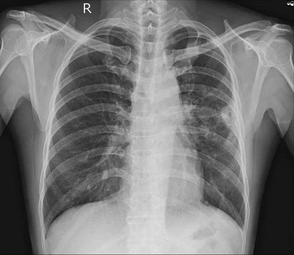
Doctor Diagnoses Pneumonia, X-ray Of Man's Lungs Isolated On White Background, Picture Of Human Lungs.
Image, 2.65MB, 4592 × 2584 jpg

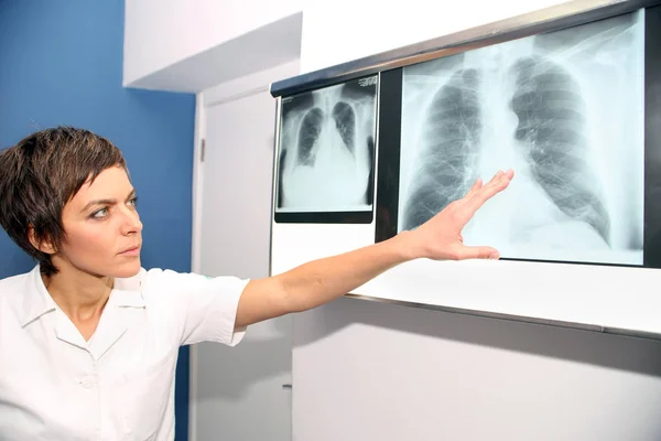
A Doctor Reads A Chest X-ray In The Bright Hospital's Waiting Room. The Concept Of Pneumonia, Respiratory Diseases. Copy Space. Banner.
Image, 6.74MB, 7573 × 3856 jpg


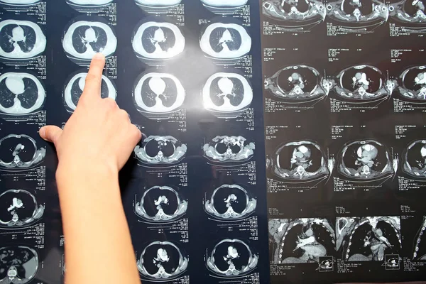


Close Up Hand Doctor Point On Computed Tomography Of The Lungs With Pneumonia Or Coronavirus Disease.High Resolution Coronavirus Covid19 Concept.
Image, 4.86MB, 4657 × 3209 jpg


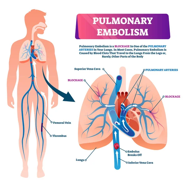
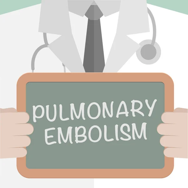
Pulmonary Embolism Disease. Consultation Or Appointment With Doctor In Clinic. Deep Vein Thrombosis. Blood Clot In Lung Vein. Blocking Blood Flow, Circulatory System Problem. DVT Vector Illustration
Vector, 0.45MB, 5074 × 4194 eps


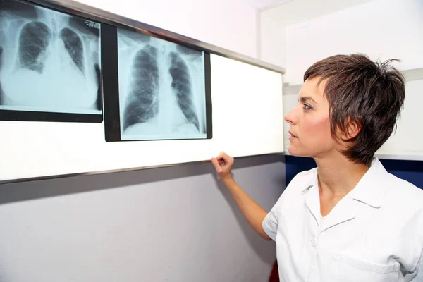
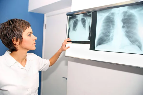
Medical Stethoscope And X-ray Or Roentgen Image. Close-up Shot Of Lung Radiography
Image, 5.91MB, 5184 × 3456 jpg

Pulmonary Embolism Symptoms. Deep Vein Thrombosis Disease. Blood Clot In Lung Vein Anatomical Poster. Blocking Blood Flow In Lungs. Circulatory System Problem. DVT Medical Flat Vector Illustration.
Vector, 0.52MB, 5336 × 3424 eps
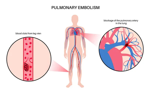
Pulmonary Embolism Disease. Deep Vein Thrombosis In Male Body. Blood Clot In Lung Vein Anatomical Poster. Blocking Blood Flow In Lungs. Circulatory System Problem. DVT Medical Flat Vector Illustration
Vector, 0.47MB, 5039 × 3647 eps

CT Scan Chest Infiltrative Mass And Nodules At Lt Upper Lobe, And Superior Segment Of Lt Lower Lobe, CA Lung Is Likely.Image Too Blurry When Views Full Solusion.
Image, 2.62MB, 3300 × 3000 jpg
Pulmonary Embolism Disease. Deep Vein Thrombosis In Male Body. Blood Clot In Lung Vein Anatomical Poster. Blocking Blood Flow In Lungs. Circulatory System Problem. DVT Medical Flat Vector Illustration
Vector, 0.73MB, 5985 × 3687 eps
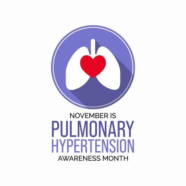
Vector Illustration On The Theme Of Pulmonary Hypertension Awareness Month Observed Each Year During November.
Vector, 5.81MB, 4000 × 4000 eps
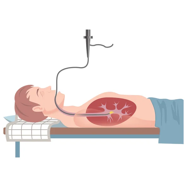
Bronchoscopy. The Patient Is In The Supine Position. Medical Examination. Vector Flat Illustration
Vector, 7.2MB, 2000 × 2000 eps

Page 1 >> Next