
Stock image Qt Prolongation

Prague, Czech Republic - JULY 10 2024: Mictonorm Box Of Medication With Propiverine Hydrochloride Active Substance By APOGEPHA Arzneimittel GmbH, Used For Treatment Of Overactive Bladder And Urinary Incontinence.
Image, 2.24MB, 4032 × 3024 jpg

Prague, Czech Republic - JULY 10 2024: Mictonorm Box Of Medication With Propiverine Hydrochloride Active Substance By APOGEPHA Arzneimittel GmbH, Used For Treatment Of Overactive Bladder And Urinary Incontinence.
Image, 2.21MB, 3024 × 4032 jpg
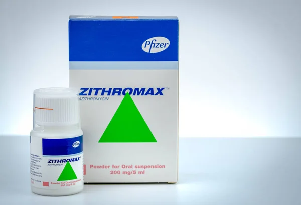
CHONBURI, THAILAND-AUGUST 3, 2018 : Zithromax Powder For Oral Suspension 200 Mg/5 Ml. Azithromycin Product Of Pfizer. Manufactured By Haupt Pharma Latina, Italy. Oral Antibiotic Drug For Infection.
Image, 10.21MB, 7777 × 5300 jpg

Prague, Czech Republic - JULY 10 2024: Mictonorm Box Of Medication With Propiverine Hydrochloride Active Substance By APOGEPHA Arzneimittel GmbH, Used For Treatment Of Overactive Bladder And Urinary Incontinence.
Image, 2.25MB, 4032 × 3024 jpg

Prague, Czech Republic - JULY 10 2024: Mictonorm Box Of Medication With Propiverine Hydrochloride Active Substance By APOGEPHA Arzneimittel GmbH, Used For Treatment Of Overactive Bladder And Urinary Incontinence.
Image, 2.22MB, 4032 × 3024 jpg

Prague, Czech Republic - JULY 10 2024: Mictonorm Box Of Medication With Propiverine Hydrochloride Active Substance By APOGEPHA Arzneimittel GmbH, Used For Treatment Of Overactive Bladder And Urinary Incontinence.
Image, 2.21MB, 3024 × 4032 jpg

CHONBURI, THAILAND-AUGUST 3, 2018 : Zithromax Powder For Oral Suspension 200 Mg/5 Ml. Azithromycin Product Of Pfizer. Manufactured By Haupt Pharma Latina, Italy. Oral Antibiotic Drug For Infection.
Image, 14.21MB, 7708 × 4813 jpg

Prague, Czech Republic - JULY 10 2024: Mictonorm Box Of Medication With Propiverine Hydrochloride Active Substance By APOGEPHA Arzneimittel GmbH, Used For Treatment Of Overactive Bladder And Urinary Incontinence.
Image, 2.18MB, 3024 × 4032 jpg

Prague, Czech Republic - JULY 10 2024: Mictonorm Box Of Medication With Propiverine Hydrochloride Active Substance By APOGEPHA Arzneimittel GmbH, Used For Treatment Of Overactive Bladder And Urinary Incontinence.
Image, 2.16MB, 3024 × 4032 jpg

Prague, Czech Republic - JULY 10 2024: Mictonorm Box Of Medication With Propiverine Hydrochloride Active Substance By APOGEPHA Arzneimittel GmbH, Used For Treatment Of Overactive Bladder And Urinary Incontinence.
Image, 2.2MB, 3024 × 4032 jpg
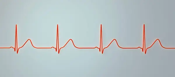
3D Illustration Of An Electrocardiogram (ECG) Showing Prolonged QT Interval With Broad-based T-waves, Characteristic Of Type 1 Long QT Syndrome.
Image, 1.22MB, 9000 × 4000 jpg

Long QT Syndrome, Type 2. 3D Illustration Of An Electrocardiogram (ECG) Showing Prolonged QT Interval With Notched And Of Lower Amplitude T-waves.
Image, 0.98MB, 7000 × 3647 jpg

Long QT Syndrome, Type 2. 3D Illustration Of An Electrocardiogram (ECG) Showing Prolonged QT Interval With Notched And Of Lower Amplitude T-waves.
Image, 1.09MB, 9000 × 4000 jpg

Long QT Syndrome, Type 2. 3D Illustration Of An Electrocardiogram (ECG) Showing Prolonged QT Interval With Notched And Of Lower Amplitude T-waves.
Image, 0.88MB, 9000 × 4000 jpg

Long QT Syndrome, Type 2. 3D Illustration Of An Electrocardiogram (ECG) Showing Prolonged QT Interval With Notched And Of Lower Amplitude T-waves.
Image, 3.79MB, 9000 × 4000 jpg

Long QT Syndrome, Type 2. 3D Illustration Of An Electrocardiogram (ECG) Showing Prolonged QT Interval With Notched And Of Lower Amplitude T-waves.
Image, 0.64MB, 7000 × 2867 jpg

Long QT Syndrome, Type 2. 3D Illustration Of An Electrocardiogram (ECG) Showing Prolonged QT Interval With Notched And Of Lower Amplitude T-waves.
Image, 5.51MB, 9000 × 4000 jpg

Long QT Syndrome, Type 2. 3D Illustration Of An Electrocardiogram (ECG) Showing Prolonged QT Interval With Notched And Of Lower Amplitude T-waves.
Image, 0.64MB, 7000 × 2867 jpg

Long QT Syndrome, Type 2. 3D Illustration Of An Electrocardiogram (ECG) Showing Prolonged QT Interval With Notched And Of Lower Amplitude T-waves.
Image, 7.28MB, 8000 × 4000 jpg

3D Illustration Of An Electrocardiogram (ECG) Showing Prolonged QT Interval With Broad-based T-waves, Characteristic Of Type 1 Long QT Syndrome.
Image, 0.96MB, 9000 × 4000 jpg

3D Illustration Of An Electrocardiogram (ECG) Showing Prolonged QT Interval With Broad-based T-waves, Characteristic Of Type 1 Long QT Syndrome.
Image, 0.71MB, 7000 × 2926 jpg

3D Illustration Of An Electrocardiogram (ECG) Showing Prolonged QT Interval With Broad-based T-waves, Characteristic Of Type 1 Long QT Syndrome.
Image, 5.58MB, 7000 × 3938 jpg

3D Illustration Of An Electrocardiogram (ECG) Showing Prolonged QT Interval With Broad-based T-waves, Characteristic Of Type 1 Long QT Syndrome.
Image, 0.71MB, 7000 × 2926 jpg

3D Illustration Of An Electrocardiogram (ECG) Showing Prolonged QT Interval With Broad-based T-waves, Characteristic Of Type 1 Long QT Syndrome.
Image, 1MB, 7000 × 3646 jpg

3D Illustration Of An Electrocardiogram (ECG) Showing Prolonged QT Interval With Broad-based T-waves, Characteristic Of Type 1 Long QT Syndrome.
Image, 5.18MB, 9000 × 4000 jpg

3D Illustration Of An Electrocardiogram (ECG) Showing Prolonged QT Interval With Broad-based T-waves, Characteristic Of Type 1 Long QT Syndrome.
Image, 5.55MB, 9000 × 4000 jpg

3D Illustration Of An Electrocardiogram (ECG) Showing Prolonged QT Interval With Broad-based T-waves, Characteristic Of Type 1 Long QT Syndrome.
Image, 1.31MB, 8000 × 4000 jpg

Long QT Syndrome, Type 2. 3D Illustration Of An Electrocardiogram (ECG) Showing Prolonged QT Interval With Notched And Of Lower Amplitude T-waves.
Image, 1.24MB, 8000 × 4000 jpg

Long QT Syndrome, Type 2. 3D Illustration Of An Electrocardiogram (ECG) Showing Prolonged QT Interval With Notched And Of Lower Amplitude T-waves.
Image, 0.95MB, 9000 × 4000 jpg

3D Illustration Of An Electrocardiogram (ECG) Showing Prolonged QT Interval With Broad-based T-waves, Characteristic Of Type 1 Long QT Syndrome.
Image, 3.59MB, 8000 × 4000 jpg

Long QT Syndrome, Type 2. 3D Illustration Of An Electrocardiogram (ECG) Showing Prolonged QT Interval With Notched And Of Lower Amplitude T-waves.
Image, 5.18MB, 7000 × 3647 jpg

Long QT Syndrome, Type 2. 3D Illustration Of An Electrocardiogram (ECG) Showing Prolonged QT Interval With Notched And Of Lower Amplitude T-waves.
Image, 5.11MB, 9000 × 4000 jpg

Long QT Syndrome, Type 2. 3D Illustration Of An Electrocardiogram (ECG) Showing Prolonged QT Interval With Notched And Of Lower Amplitude T-waves.
Image, 3.89MB, 7000 × 2891 jpg

3D Illustration Of An Electrocardiogram (ECG) Showing Prolonged QT Interval With Broad-based T-waves, Characteristic Of Type 1 Long QT Syndrome.
Image, 2.83MB, 7000 × 3500 jpg

Long QT Syndrome, Type 2. 3D Illustration Of An Electrocardiogram (ECG) Showing Prolonged QT Interval With Notched And Of Lower Amplitude T-waves.
Image, 2.79MB, 7000 × 3647 jpg

Long QT Syndrome, Type 2. 3D Illustration Of An Electrocardiogram (ECG) Showing Prolonged QT Interval With Notched And Of Lower Amplitude T-waves.
Image, 5.22MB, 9000 × 4000 jpg

Long QT Syndrome, Type 2. 3D Illustration Of An Electrocardiogram (ECG) Showing Prolonged QT Interval With Notched And Of Lower Amplitude T-waves.
Image, 1.97MB, 9000 × 4000 jpg

3D Illustration Of An Electrocardiogram (ECG) Showing Prolonged QT Interval With Broad-based T-waves, Characteristic Of Type 1 Long QT Syndrome.
Image, 1.13MB, 9000 × 4000 jpg

Long QT Syndrome, Type 2. 3D Illustration Of An Electrocardiogram (ECG) Showing Prolonged QT Interval With Notched And Of Lower Amplitude T-waves.
Image, 1.17MB, 9000 × 4000 jpg

3D Illustration Of An Electrocardiogram (ECG) Showing Prolonged QT Interval With Broad-based T-waves, Characteristic Of Type 1 Long QT Syndrome.
Image, 0.96MB, 9000 × 4000 jpg

3D Illustration Of An Electrocardiogram (ECG) Showing Prolonged QT Interval With Broad-based T-waves, Characteristic Of Type 1 Long QT Syndrome.
Image, 3.59MB, 8000 × 4000 jpg

3D Illustration Of An Electrocardiogram (ECG) Showing Prolonged QT Interval With Broad-based T-waves, Characteristic Of Type 1 Long QT Syndrome.
Image, 1.03MB, 9000 × 4000 jpg

Long QT Syndrome, Type 2. 3D Illustration Of An Electrocardiogram (ECG) Showing Prolonged QT Interval With Notched And Of Lower Amplitude T-waves.
Image, 2.79MB, 7000 × 3647 jpg

ECG Displaying Torsades De Pointes Rhythm, Dangerous Heart Rhythm With Fast, Irregular Beats Twisting Around The Electrical Axis, Potentially Causing Fainting Or Cardiac Arrest, 3D Illustration.
Image, 9.87MB, 9000 × 6000 jpg

Normal T Waves Are Asymmetrical. Coronary T Waves Are Not Absolutely Symmetric, But Increase In Symmetry. From The Bottom Of The T Wave To The Bottom, The Asymmetry Gradually Increases.
Image, 6.72MB, 11822 × 8404 jpg

3D Illustration Of An Electrocardiogram (ECG) Showing Prolonged QT Interval With Broad-based T-waves, Characteristic Of Type 1 Long QT Syndrome.
Image, 4.63MB, 6420 × 3210 jpg

Long QT Syndrome, Type 2. 3D Illustration Of An Electrocardiogram (ECG) Showing Prolonged QT Interval With Notched And Of Lower Amplitude T-waves.
Image, 5.51MB, 9000 × 4000 jpg

Long QT Syndrome, Type 2. 3D Illustration Of An Electrocardiogram (ECG) Showing Prolonged QT Interval With Notched And Of Lower Amplitude T-waves.
Image, 3.89MB, 7000 × 2891 jpg

3D Illustration Of An Electrocardiogram (ECG) Showing Prolonged QT Interval With Broad-based T-waves, Characteristic Of Type 1 Long QT Syndrome.
Image, 3.84MB, 9000 × 4000 jpg

Long QT Syndrome, Type 2. 3D Illustration Of An Electrocardiogram (ECG) Showing Prolonged QT Interval With Notched And Of Lower Amplitude T-waves.
Image, 7.28MB, 8000 × 4000 jpg
Page 1 >> Next