
Stock image Signal Protein
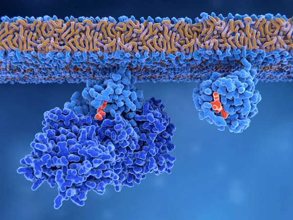
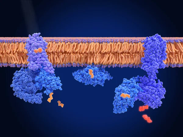
Activation Of A Ras Protein Inactive Ras Protein (left) Is Activated By A GEF Protein Opening The Binding Site Allowing GDP To Exit. Then GTP Can Bind To RAS Turning It Into The Active Form. 3d Render. Illustration
Image, 3.68MB, 8000 × 6000 jpg

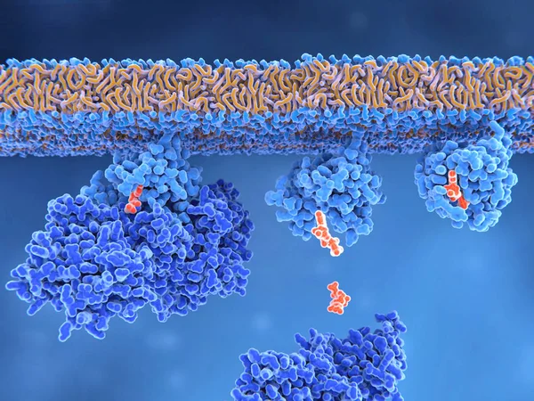
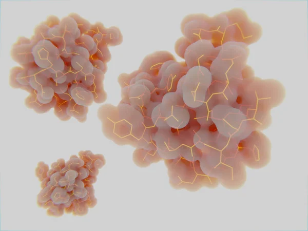
3d Computer Illustration Of An Activated Ras Protein With GTP Bound. Ras Proteins Are Involved In Transmitting Signals Within Cells Turning On Genes Involved In Cell Growth, Differentiation And Survival. Mutations In Ras Genes Can Lead To Permanentl
Image, 1.5MB, 8000 × 6000 jpg
3d Computer Illustration Of The Activation Process Of A Ras Protein. Inactive Ras Protein (left) Is Activated By A GEF Protein Opening The Binding Site And Allowing GDP To Exit. Afterwards GTP Can Bind To RAS Turning It Into The Active Form (right).
Image, 7.61MB, 8000 × 6000 jpg
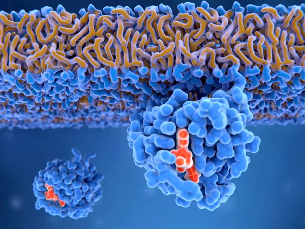
3d Computer Illustration Of An Activated Ras Protein. Ras Proteins Are Involved In Transmitting Signals Within Cells Turning On Genes Involved In Cell Growth, Differentiation And Survival. Mutations In Ras Genes Can Lead To Permanently Activated Prot
Image, 4.61MB, 8000 × 6000 jpg
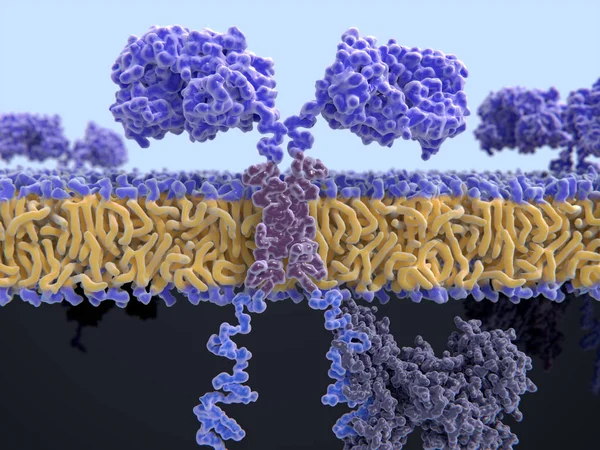
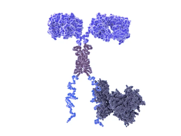
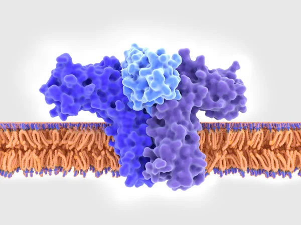
3d Computer Illustration Of A Chimeric Antigen Receptor. CARs Are Engineered Cell Receptors That Allow T Cells To Recognize And Attack Cancer Cells In A Specific Way. They Are Built By Connecting Several Functional Parts From Different Proteins.
Image, 8.45MB, 8000 × 6000 jpg
3d Computer Illustration Of A Chimeric Antigen Receptor. CARs Are Engineered Cell Receptors That Allow T Cells To Recognize/attack Specifically Cancer Cells. A Signal Protein Is Attached To The Intracellular Domain.
Image, 3.45MB, 8000 × 6000 jpg
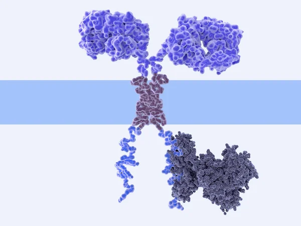
3d Computer Illustration Of A Chimeric Antigen Receptor. CARs Are Engineered Cell Receptors That Allow T Cells To Recognize/attack Specifically Cancer Cells. A Signal Protein Is Attached To The Intracellular Domain.
Image, 2.19MB, 8000 × 6000 jpg

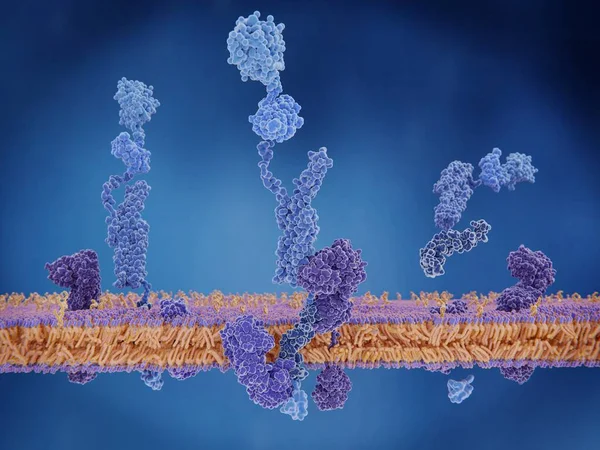
T-cell Receptors Are Similar To One Arm Of An Antibody. Like Antibodies, They Are Composed Of Two Chains. The Binding Site Is At The Tip Of The Molecule,
Image, 2.5MB, 8000 × 6000 jpg
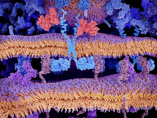
Engineered Receptors (light Blue) On The Surface Of A T-lymphocyte Bind Specifically To CD19-antigen Molecules (magenta) On A Leukemia Cell. This Activates A Signal Cascade In The T-cell Leading To The Segregation Of Vesicles That Contain Perforin An
Image, 11.44MB, 8000 × 6000 jpg
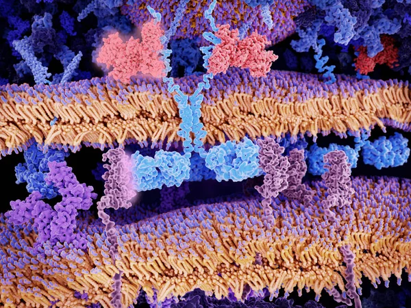
Engineered Receptors (light Blue) On The Surface Of A T-lymphocyte Bind Specifically To CD19-antigen Molecules (magenta) On A Leukemia Cell. This Activates A Signal Cascade In The T-cell Leading To The Apoptosis Of The Cancer Cell.
Image, 11.66MB, 8000 × 6000 jpg
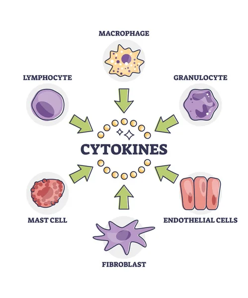
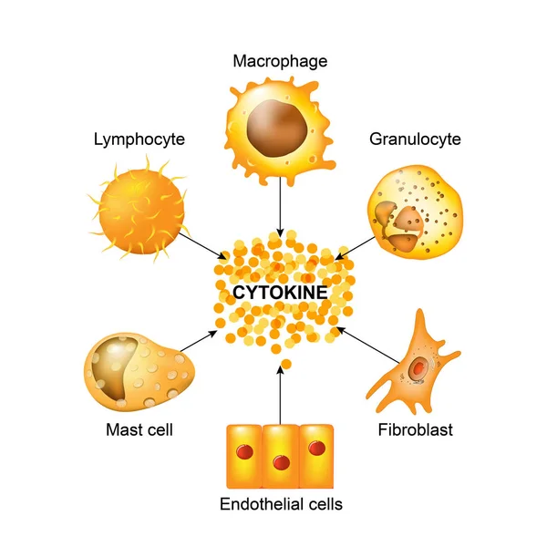
Cytokines Releasing Cells List For Immune System Response Outline Diagram. Labeled Educational Scheme With Macrophage, Granulocyte, Endothelial, Fibroblast, Mast And Lymphocyte Vector Illustration.
Vector, 5.64MB, 4000 × 4800 eps

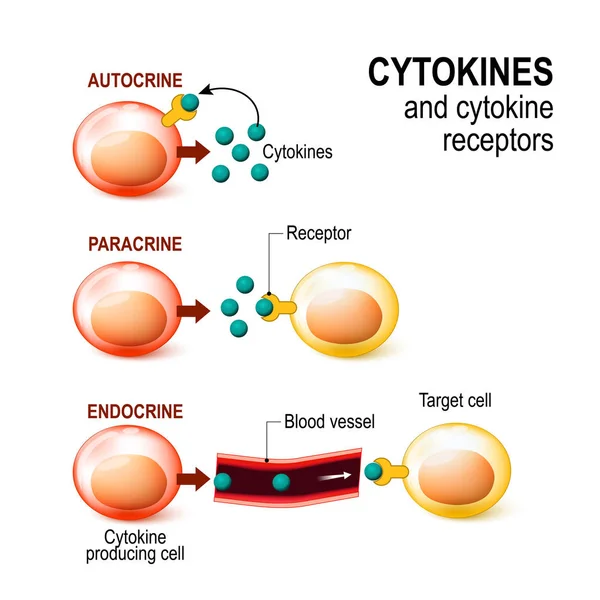
Hormones, Receptors And Target Cells. Each Type Of Hormone Is Designed Only Certain Cells. These Cells Will Have Receptors On Them That Are Specific For A Certain Hormone. Vector Illustration For Medical, Biological, And Educational Use
Vector, 2.77MB, 5013 × 5012 eps

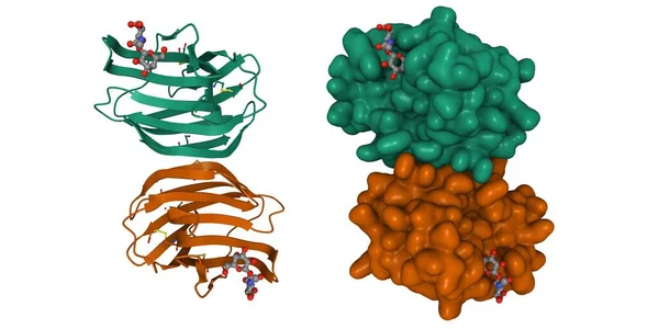
Structure Of Human Hormone Insulin-like Peptide-5 Heterodimer, 3D Cartoon And Gaussian Surface Models, White Background
Image, 2.56MB, 8000 × 4000 jpg
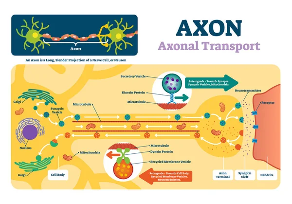
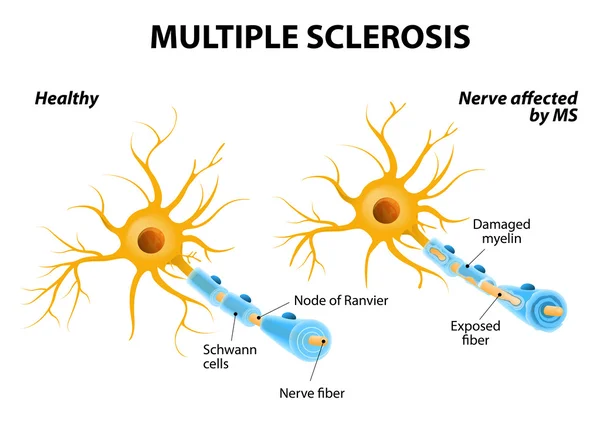
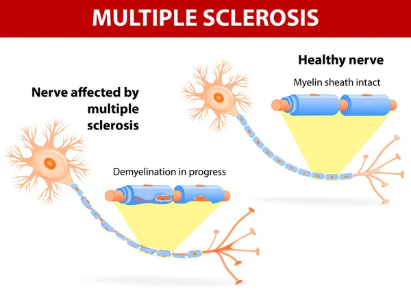
Axon Vector Illustration. Labeled Diagram With Explanation And Structure.
Vector, 5.97MB, 5000 × 3498 eps

Neural Synapses, Failure In Their Functioning Causes Degenerative Neurological Diseases Such As Alzheimer's, Parkson's And Dementia. 3D Rendering
Image, 2.96MB, 3508 × 2480 jpg
Structure Of Follicle-stimulating Hormone (color) In Complex With The Entire Ectodomain Of Its Receptor (grey), 3D Gaussian Surface Model, White Background
Image, 0.75MB, 6000 × 4000 jpg
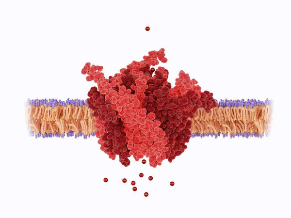
The Calcium Channel Is Composed Of A Hexameric Assembly Or Orai Subunits Around A Central Ion Pore. The Channel Shows Selective Permeability To Calcium Ions.
Image, 3.65MB, 8000 × 6000 jpg

Dopamine And The Dopamine Receptor D1. The Dopamine Binding Side Is Insinuated. Source: PDB Entry 7ljd
Image, 6.96MB, 8000 × 6000 jpg


Integrin Alpha2 I Domain (green) In Complex With Collagen, 3D Cartoon Model, White Background
Image, 2.52MB, 6000 × 4000 jpg


Calmodulin, Inactive-calcium Free (left), And Activated (right) Conformations,
Image, 2.57MB, 8000 × 6000 jpg
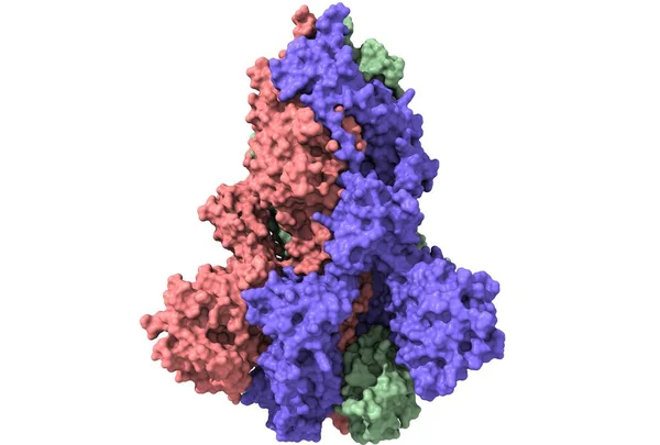
The Amyloid Precursor Protein. When Cleaved, The Membrane Domain Is Involved In The Alzheimer Disease Building Amyloid Plaques. 3d Rendering. Illustration
Image, 3.29MB, 8000 × 6000 jpg

Activation Of The GABA B Receptor By Baclofen. GABA B Receptors Are G Protein-coupled Receptors. Binding Of An Agonist (baclofen, Red) Leads To A G-protein Coupled C-AMP Signaling Pathway. Source: PDB Entries 7eb2, 6r3q,.
Image, 9.97MB, 8000 × 6000 jpg

Rhodopsin Is A Light Sensitive G-protein Coupled Receptor With Retinal As Cofactor. That Stimulates The G-protein Transducin, Resulting In The Liberation Of Its Subunit. This GTP-bound Subunit In Turn Activates CGMP Phosphodiesterase.
Image, 8.93MB, 8000 × 6000 jpg
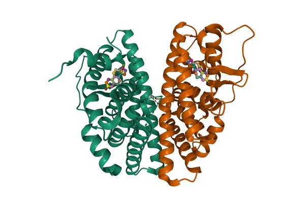
Estrogen Receptor Beta Dimer In Complex With Estradiol, 3D Cartoon Model, Chain Id Color Scheme, Based On PDB 5toa, White Background
Image, 2.91MB, 6000 × 4000 jpg
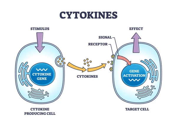
Cytokines Process As Micro Proteins For Cell Signaling Outline Diagram. Labeled Educational Scheme With Immune Response And Producing Or Target Cells Vector Illustration. Stimulus And Antibody Effect.
Vector, 5.6MB, 5000 × 3571 eps


Structure Of Human Hormone Insulin-like Peptide-3 Heterodimer, 3D Cartoon And Gaussian Surface Models, White Background
Image, 3.16MB, 10000 × 4000 jpg

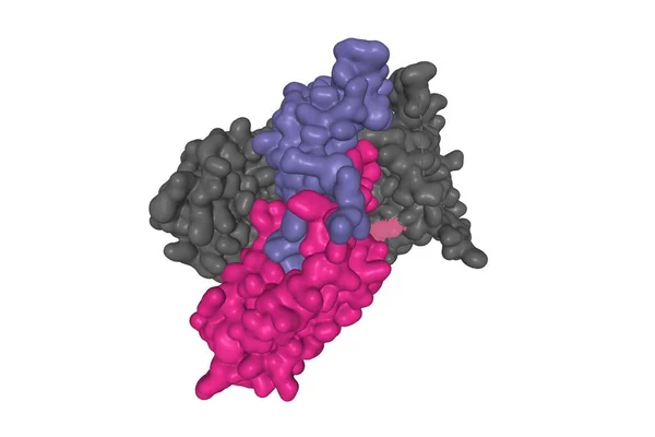
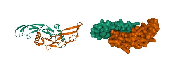
Crystal Structure Of Human Galectin-1 In Complex With Type 1 N-acetyllactosamine. 3D Cartoon And Gaussian Surface Model, Chain Id Color Scheme, PDB 4xbl, White Background
Image, 3.8MB, 8000 × 4000 jpg
Structure Of Bone Morphogenetic Protein 3 Homodimer, 3D Cartoon And Gaussian Surface Model, White Background
Image, 3.12MB, 10000 × 4100 jpg
Structure Of The SARS-CoV-2 Spike Glycoprotein, Surface Model, White Background, 3D Illustration Isolated
Image, 0.81MB, 5000 × 3380 jpg
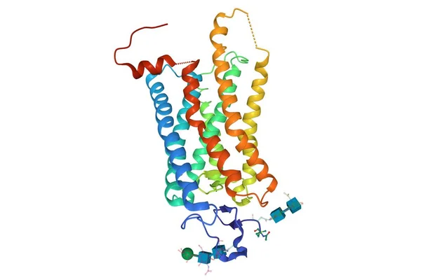
Crystal Structure Of A Photoactivated Rhodopsin, 3D Cartoon Model Isolated, White Background
Image, 1.92MB, 6000 × 4000 jpg


Calmodulin, A Crucial Messenger Protein. Calmodulin Has 4 Ca2+ Binding Sites.
Image, 2.97MB, 8000 × 6000 jpg


CAMP Cyclic Adenosine MonoPhosphate - Second Messenger Important In Many Biological Processes, Acronym Text On Blackboard
Image, 11.5MB, 5760 × 3840 jpg
Page 1 >> Next