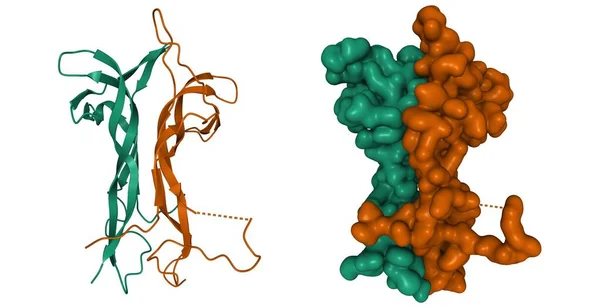
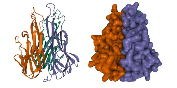
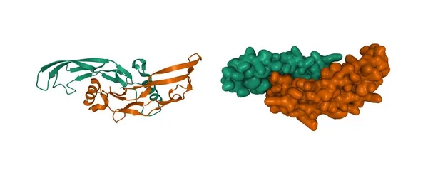
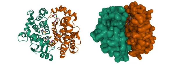
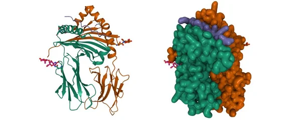
Stock image Crystal structure of human galectin-1 in complex with type 1 N-acetyllactosamine. 3D cartoon and Gaussian surface model, chain id color scheme, PDB 4xbl, white background
Published: Oct.26, 2021 08:18:35
Author: unnaugan
Views: 6
Downloads: 2
File type: image / jpg
File size: 3.8 MB
Orginal size: 8000 x 4000 px
Available sizes:
Level: beginner