photo free
Home Images Vectors Videos

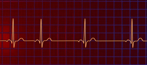
Stock image Sinus Tachycardia

stock vector Schemes set of common electrocardiogram (ECG) abnormalities, including partial blocks and flutter

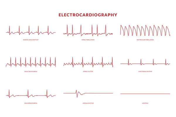
Schemes Set Of Common Electrocardiogram (ECG) Abnormalities, Including Partial Blocks And Flutter

Vector, 9.68MB, 7750 × 4367 eps
stock vector Electrocardiography Heartbeat Line monitor. vector EPS10 Illustration

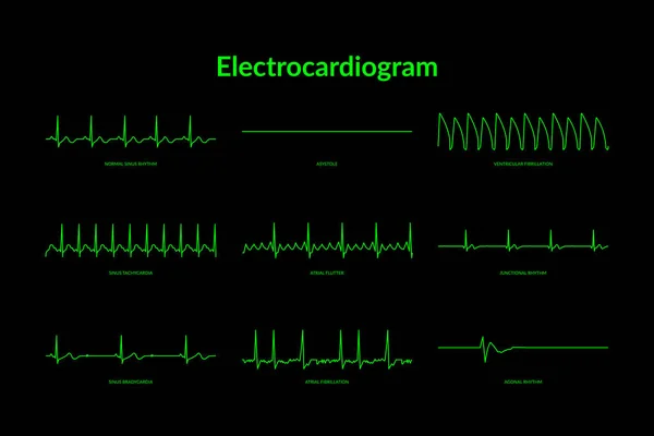
Electrocardiography Heartbeat Line Monitor. Vector EPS10 Illustration

Vector, 0.82MB, 6000 × 4000 eps
stock image Heart rate on paper for recording an electrocardiogram, prevention of heart diseases. Electrocardiogram strips with cardiac arrhythmias. Alterations of heartbeats represented on paper. Copy space.

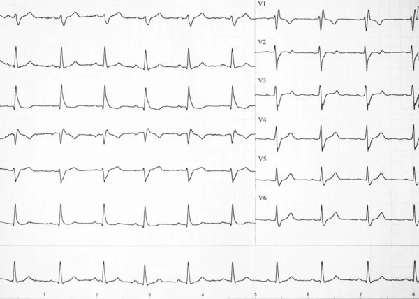
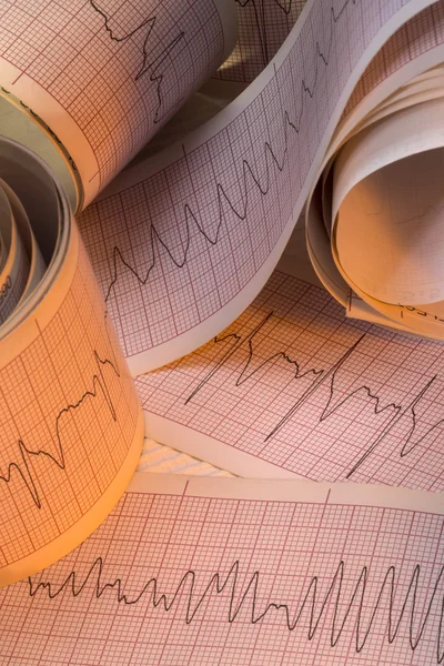
Heart Rate On Paper For Recording An Electrocardiogram, Prevention Of Heart Diseases. Electrocardiogram Strips With Cardiac Arrhythmias. Alterations Of Heartbeats Represented On Paper. Copy Space.

Image, 3.11MB, 4675 × 3339 jpg
stock image Young patient at the reception of a doctor cardiologist-psychotherapist. The concept of psychological disorder and heart pain, cardiac neurosis, cardiophobia, psychotherapy, psychosomatics

Young Patient At The Reception Of A Doctor Cardiologist-psychotherapist. The Concept Of Psychological Disorder And Heart Pain, Cardiac Neurosis, Cardiophobia, Psychotherapy, Psychosomatics

Image, 2.49MB, 5376 × 3444 jpg
stock vector ECG Heartbeat Line. Electrocardiogram vector illustration. Sinus Tachycardia

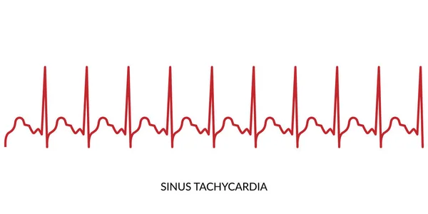
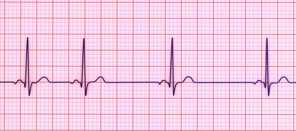
ECG Heartbeat Line. Electrocardiogram Vector Illustration. Sinus Tachycardia

Vector, 0.58MB, 5600 × 2800 eps
stock vector Electrocardiogram Heartbeat Line monitor. vector EPS10 Illustration

Electrocardiogram Heartbeat Line Monitor. Vector EPS10 Illustration

Vector, 0.82MB, 6000 × 4000 eps
stock vector Types of cardiac arrhythmia: sinus tachycardia, sinus arrhythmia

Types Of Cardiac Arrhythmia: Sinus Tachycardia, Sinus Arrhythmia

Vector, 5.31MB, 5315 × 5315 eps
stock image A 2:1 left bundle branch block is considered when complete left bundle branch block alternates with normal QRS complexes and the PR interval is fixed.

A 2:1 Left Bundle Branch Block Is Considered When Complete Left Bundle Branch Block Alternates With Normal QRS Complexes And The PR Interval Is Fixed.

Image, 5.72MB, 10000 × 3162 jpg
stock image Tachycardia. Medicine. 3D Illustration.

Tachycardia. Medicine. 3D Illustration.

Image, 3.38MB, 5000 × 3300 jpg
stock vector Vector medical illustration with emblem of tachycardia

Vector Medical Illustration With Emblem Of Tachycardia

Vector, 2.17MB, 4167 × 4167 eps
stock image Arrhythmia. Medicine. 3D Illustration.

Arrhythmia. Medicine. 3D Illustration.

Image, 3.43MB, 5000 × 3300 jpg
stock image Arrhythmia Diagnosis. Medical Concept.

Arrhythmia Diagnosis. Medical Concept.

Image, 9.02MB, 5733 × 3780 jpg
stock image Bidirectional ventricular tachycardia is a kind of malignant arrhythmia. The polarity of QRS main wave alternates from beat to beat, and it is easy to degenerate into ventricular fibrillation.

Bidirectional Ventricular Tachycardia Is A Kind Of Malignant Arrhythmia. The Polarity Of QRS Main Wave Alternates From Beat To Beat, And It Is Easy To Degenerate Into Ventricular Fibrillation.

Image, 10.66MB, 10000 × 4450 jpg
stock image Arrhythmia - Phrase in Differential Diagnoses. 3D Render.

Arrhythmia - Phrase In Differential Diagnoses. 3D Render.

Image, 11.97MB, 5000 × 2813 jpg
stock image Arrhythmia - Inscription in Anamnesis. 3D Render.

Arrhythmia - Inscription In Anamnesis. 3D Render.

Image, 12.03MB, 5000 × 2813 jpg
stock image Diagnosis - Arrhythmia. Medical Concept. 3D Render.

Diagnosis - Arrhythmia. Medical Concept. 3D Render.

Image, 8.79MB, 5733 × 3780 jpg
stock image The illustration shows the two patterns of ventricular tachycardia episodes.The green circle represents sinus rhythm. Picture A shows paroxysmal episodes of ventricular tachycardia, and picture B shows short bursts.

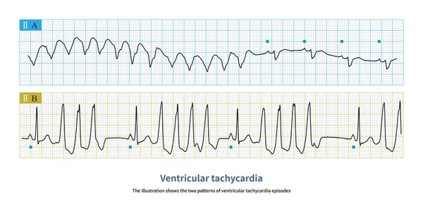
The Illustration Shows The Two Patterns Of Ventricular Tachycardia Episodes.The Green Circle Represents Sinus Rhythm. Picture A Shows Paroxysmal Episodes Of Ventricular Tachycardia, And Picture B Shows Short Bursts.

Image, 10.72MB, 10000 × 5059 jpg
stock image EKG, Pulse, BP and Respiration ICU Monitor Screen

EKG, Pulse, BP And Respiration ICU Monitor Screen

Image, 1.84MB, 3264 × 2448 jpg
stock vector heartbeat icon. ECG Pathology Trace

Heartbeat Icon. ECG Pathology Trace

Vector, 1.02MB, 6000 × 4000 eps
stock image Arrhythmias - Wording in Anamnesis. 3D Render.

Arrhythmias - Wording In Anamnesis. 3D Render.

Image, 11.99MB, 5000 × 2813 jpg
stock vector heartbeat icon. ECG Pathology Trace

Heartbeat Icon. ECG Pathology Trace

Vector, 0.99MB, 6667 × 4000 eps
stock image Coffee or caffeine and heart arrhythmias (irregular heartbeat). Stethoscope and ECG tape on background of coffee beans. Effect and risk of drinking coffee or caffeine on cardiac arrhythmia development

Coffee Or Caffeine And Heart Arrhythmias (irregular Heartbeat). Stethoscope And ECG Tape On Background Of Coffee Beans. Effect And Risk Of Drinking Coffee Or Caffeine On Cardiac Arrhythmia Development

Image, 9.64MB, 6000 × 4000 jpg
stock image Arrhythmia. Medical Concept on Green Background.

Arrhythmia. Medical Concept On Green Background.

Image, 4.97MB, 5000 × 3298 jpg
stock image ECG curve folded on paper

ECG Curve Folded On Paper

Image, 2.82MB, 3956 × 3000 jpg
stock image EKG, Pulse, BP and Respiration ICU Monitor Screen

EKG, Pulse, BP And Respiration ICU Monitor Screen

Image, 2.24MB, 4032 × 2268 jpg
stock vector Full page of EKG strips with title and spot illustrations

Full Page Of EKG Strips With Title And Spot Illustrations

Vector, 5.44MB, 8164 × 5899 ai
stock vector Cardiogram, normal heart rhythm

Cardiogram, Normal Heart Rhythm

Vector, 5.79MB, 6392 × 3505 eps
stock image EKG, Pulse, BP and Respiration ICU Monitor Screen

EKG, Pulse, BP And Respiration ICU Monitor Screen

Image, 2.62MB, 3264 × 2448 jpg
stock image Electrocardiograph Traces - Cardiac Arrhythmia

Electrocardiograph Traces - Cardiac Arrhythmia

Image, 11.43MB, 3413 × 5120 jpg
stock image Young female doctor sitting at desk with medical documents, electrocardiogram record, heart ekg cardiogram chart of wave in paper in light office in hospital. Woman in medical gown in consulting room.

Young Female Doctor Sitting At Desk With Medical Documents, Electrocardiogram Record, Heart Ekg Cardiogram Chart Of Wave In Paper In Light Office In Hospital. Woman In Medical Gown In Consulting Room.

Image, 7.57MB, 5760 × 3840 jpg
stock image A 14-year-old leukemic child had a sudden wide QRS tachycardia with a frequency of 167 bpm, and the rhythm was regular. After anti-arrhythmia treatment, the patient recovered to sinus rhythm.

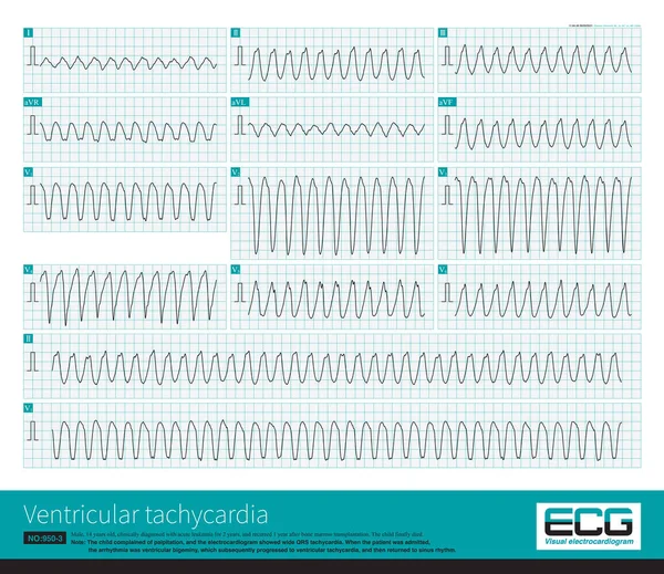
A 14-year-old Leukemic Child Had A Sudden Wide QRS Tachycardia With A Frequency Of 167 Bpm, And The Rhythm Was Regular. After Anti-arrhythmia Treatment, The Patient Recovered To Sinus Rhythm.

Image, 32.12MB, 10000 × 8649 jpg
stock image EKG, Pulse, BP and Respiration ICU Monitoring Screen

EKG, Pulse, BP And Respiration ICU Monitoring Screen

Image, 2.61MB, 4000 × 3000 jpg
stock image Ivabradine molecule. It is angina pectoris drug. Molecular model. 3D rendering. Illustration

Ivabradine Molecule. It Is Angina Pectoris Drug. Molecular Model. 3D Rendering. Illustration

Image, 4.23MB, 8000 × 3300 jpg
stock vector Ivabradine molecule. It is angina pectoris drug. Structural chemical formula and molecule model. Vector illustration

Ivabradine Molecule. It Is Angina Pectoris Drug. Structural Chemical Formula And Molecule Model. Vector Illustration

Vector, 0.42MB, 5905 × 4234 eps
stock image Ivabradine molecule. It is angina pectoris drug. Molecular model. 3D rendering. Illustration

Ivabradine Molecule. It Is Angina Pectoris Drug. Molecular Model. 3D Rendering. Illustration

Image, 2.64MB, 5070 × 3380 jpg
stock image EKG, Pulse, BP and Respiration ICU Monitor - multiple patients

EKG, Pulse, BP And Respiration ICU Monitor - Multiple Patients

Image, 2.5MB, 3860 × 2510 jpg
stock vector Ivabradine molecule. It is angina pectoris drug. Molecule model. Sheet of paper in a cage. Vector illustration

Ivabradine Molecule. It Is Angina Pectoris Drug. Molecule Model. Sheet Of Paper In A Cage. Vector Illustration

Vector, 0.46MB, 5916 × 4226 eps
stock image A detailed 3D illustration of an Electrocardiogram ECG displaying sinus arrhythmia, a condition characterized by irregular heart rhythms originating from the sinus node.

A Detailed 3D Illustration Of An Electrocardiogram ECG Displaying Sinus Arrhythmia, A Condition Characterized By Irregular Heart Rhythms Originating From The Sinus Node.

Image, 12.85MB, 9000 × 4000 jpg
stock vector Ivabradine molecule. It is angina pectoris drug. Skeletal chemical formula. Paper packaging for drugs. Vector illustration

Ivabradine Molecule. It Is Angina Pectoris Drug. Skeletal Chemical Formula. Paper Packaging For Drugs. Vector Illustration

Vector, 0.36MB, 6502 × 3845 eps
stock image EKG, Pulse, BP and Respiration ICU Monitoring Screen

EKG, Pulse, BP And Respiration ICU Monitoring Screen

Image, 3.09MB, 4000 × 3000 jpg
stock image A detailed 3D illustration of an Electrocardiogram ECG displaying sinus arrhythmia, a condition characterized by irregular heart rhythms originating from the sinus node.

A Detailed 3D Illustration Of An Electrocardiogram ECG Displaying Sinus Arrhythmia, A Condition Characterized By Irregular Heart Rhythms Originating From The Sinus Node.

Image, 9.76MB, 9000 × 4000 jpg
stock image Ivabradine molecule. It is angina pectoris drug. Molecular model. 3D rendering. Illustration

Ivabradine Molecule. It Is Angina Pectoris Drug. Molecular Model. 3D Rendering. Illustration

Image, 7.79MB, 9400 × 5150 jpg
stock vector Ivabradine molecule. It is angina pectoris drug. Structural chemical formula, dark blue background. Vector illustration

Ivabradine Molecule. It Is Angina Pectoris Drug. Structural Chemical Formula, Dark Blue Background. Vector Illustration

Vector, 0.29MB, 6642 × 3764 eps
stock image ECG displaying Torsades de pointes rhythm, dangerous heart rhythm with fast, irregular beats twisting around the electrical axis, potentially causing fainting or cardiac arrest, 3D illustration.

ECG Displaying Torsades De Pointes Rhythm, Dangerous Heart Rhythm With Fast, Irregular Beats Twisting Around The Electrical Axis, Potentially Causing Fainting Or Cardiac Arrest, 3D Illustration.

Image, 9.87MB, 9000 × 6000 jpg
Page 1 >> Next