
Stock image Submucosa
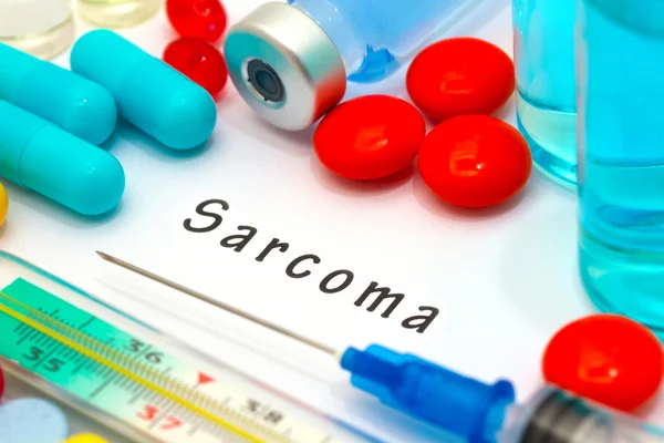
Sarcoma - Diagnosis Written On A White Piece Of Paper. Syringe And Vaccine With Drugs.
Image, 9.83MB, 5184 × 3456 jpg
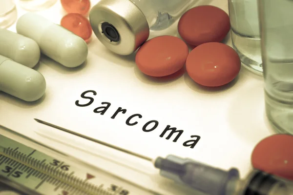
Sarcoma - Diagnosis Written On A White Piece Of Paper. Syringe And Vaccine With Drugs.
Image, 7.19MB, 4758 × 3172 jpg

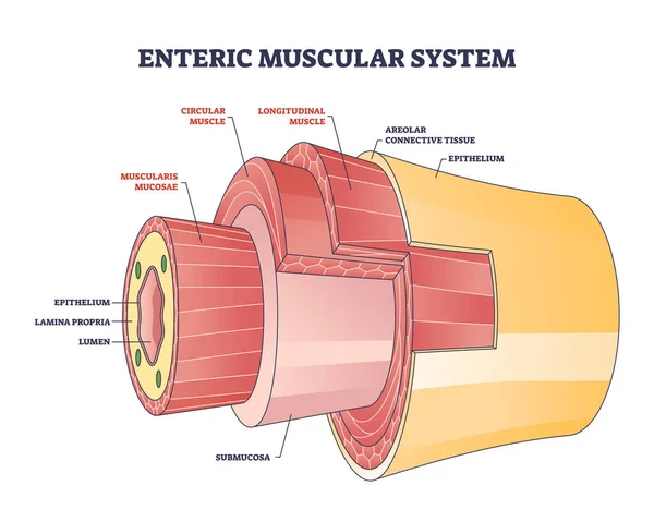
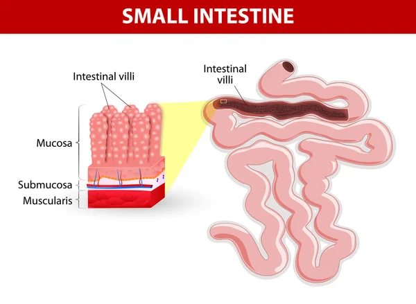
Enteric Muscular System In Gut Wall Of The Small Intestine Outline Diagram. Labeled Educational Scheme With Layers And Structure Of Digestive Tract Muscle Vector Illustration. Lamina Propria Location.
Vector, 7.06MB, 4500 × 3600 eps
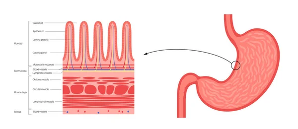
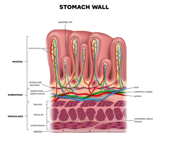
Mucous Membrane Anatomical Poster. Stomach Wall Structure. Soft Tissue That Lines The Canals And Organs In The Digestive System. Mucosa, Submucosa, Muscle Layer And Serosa Medical Vector Illustration.
Vector, 0.89MB, 6900 × 2997 eps
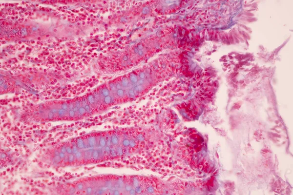
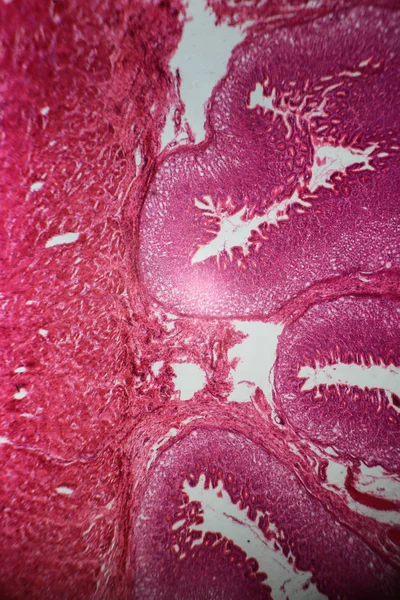
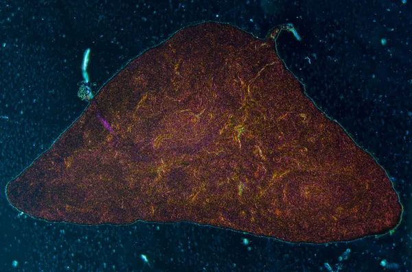
Tissue Of Small Intestine (Duodenum), Large Intestine Human And Stomach Human Under The Microscope In Lab.
Image, 17.29MB, 8192 × 5461 jpg
Tissue Of Small Intestine (Duodenum) And Vermiform Appendix Human Under The Microscope In Lab.
Image, 22.69MB, 6000 × 4000 jpg

Tissue Of Small Intestine (Duodenum) And Vermiform Appendix Human Under The Microscope In Lab.
Image, 20.31MB, 6000 × 4000 jpg

Sarcoma - Diagnosis Written On A White Piece Of Paper. Syringe And Vaccine With Drugs.
Image, 8.89MB, 5184 × 3456 jpg
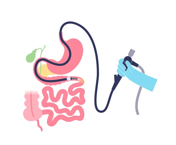
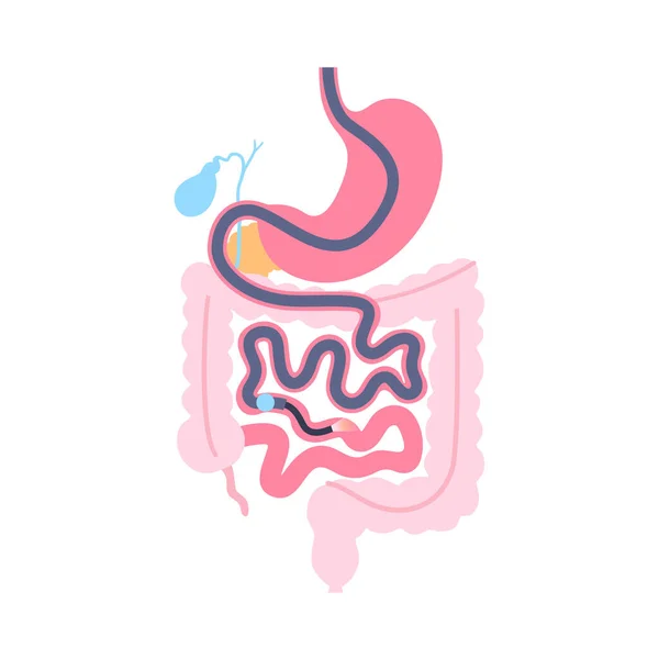
Balloon Assisted Enteroscopy. Visualization Of The Small Intestine Nonsurgical Procedure. Gastrointestinal Tract Exam. Biopsy, Polyp Removal, Bleeding Therapy Or Stent Placement Vector Illustration
Vector, 0.53MB, 4585 × 3789 eps
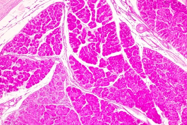
Tissue Of Small Intestine (Duodenum) And Vermiform Appendix Human Under The Microscope In Lab.
Image, 21.54MB, 6000 × 4000 jpg
Tissue Of Small Intestine (Duodenum) And Vermiform Appendix Human Under The Microscope In Lab.
Image, 18.99MB, 6000 × 4000 jpg


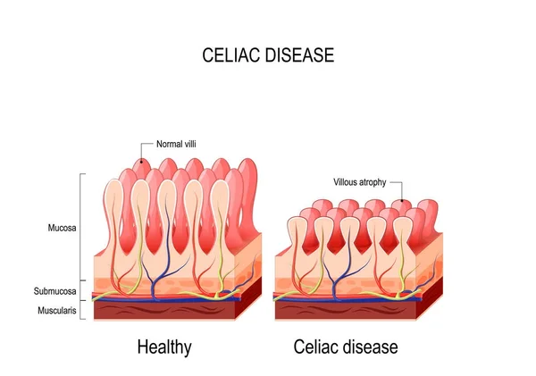
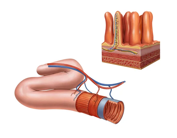
Intestinal Villus. Different Between Villi And Microvilli. Cross Section Of A Duodenum With Submucosa, Mucosa And Muscularis Layers. Vertical Section Of A Villus With Lymph Capillary Or Lacteal And Blood Vessels. Close-up Of A Columnar Epithelium, An
Vector, 2.33MB, 5000 × 3404 eps


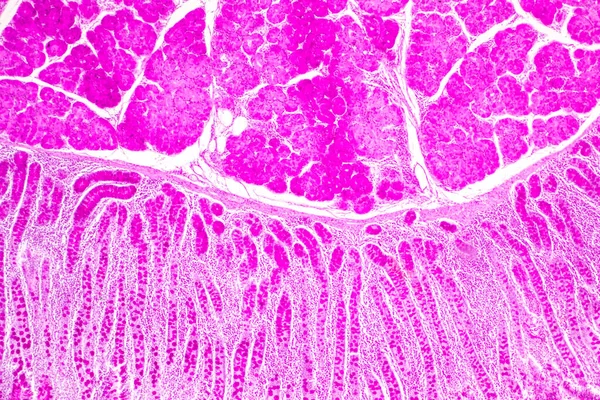
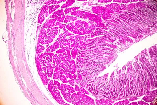
Duodenum Biopsy From The Pathology Of Small Intestine. Education For Human Histology.
Image, 13.92MB, 6000 × 4000 jpg
Single Balloon Enteroscopy Procedure. Visualization Of The Small Intestine Nonsurgical Technique. Gastrointestinal Tract Problem. Biopsy, Polyp Removal, Bleeding Therapy Or Stent Placement Flat Vector
Vector, 0.48MB, 4168 × 4167 eps

Tissue Of Small Intestine (Duodenum) And Vermiform Appendix Human Under The Microscope In Lab.
Image, 17.16MB, 6000 × 4000 jpg
Doctor's Hands In Blue Gloves Shows The Word Sarcoma. Medical Concept.
Image, 2.84MB, 3393 × 1986 jpg

Esophagogastroduodenoscopy Medical Poster. Diagnostic Endoscopic Minimally Invasive Procedure. Visualization Of The Oropharynx, Esophagus, Stomach, And Proximal Duodenum. Gastroenterology Flat Vector
Vector, 0.64MB, 5840 × 3615 eps
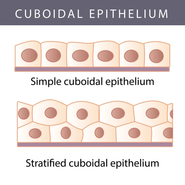
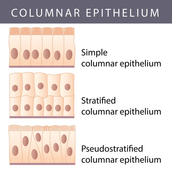
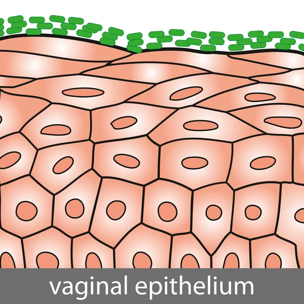
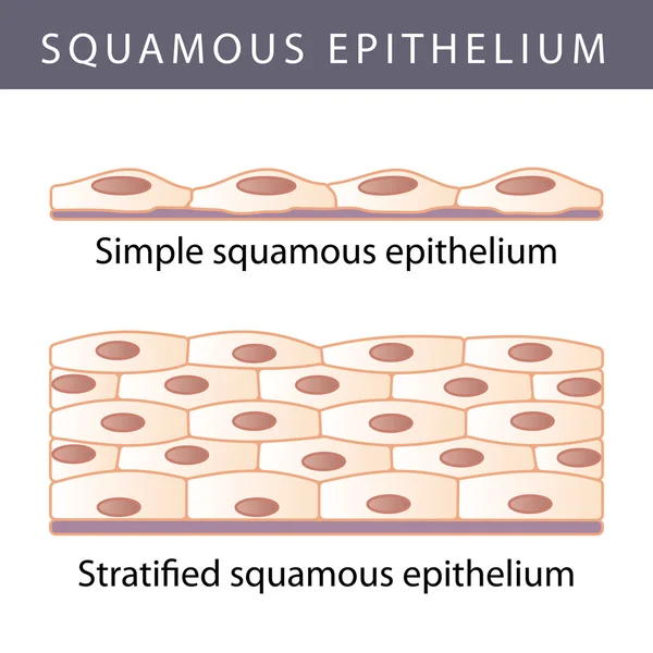
Epithelium. Squamous, Cubic, Ciliated, Glandular. Set. Infographics. Vector Illustration.
Vector, 1.56MB, 5000 × 5093 eps

The Structure Of The Glandular Epithelium. Infographics. Vector Illustration On Isolated Background
Vector, 0.78MB, 5000 × 5000 eps
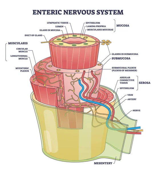
Enteric Nervous System Or ENS Intrinsic Autonomic Anatomy Outline Diagram. Labeled Educational Scheme With Complex Detailed Structure With Mucosa, Muscularis And Mesentery Part Vector Illustration.
Vector, 7.96MB, 4200 × 4600 eps
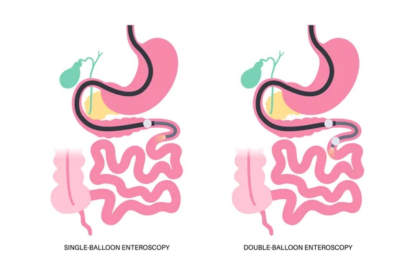
Double And Single Balloon Enteroscopy Minimally Invasive Procedure. Visualization Of The Small Intestine. Biopsy, Polyp Removal, Bleeding Therapy Or Stent Placement In Gastrointestinal Tract .poster.
Vector, 0.61MB, 5163 × 3365 eps
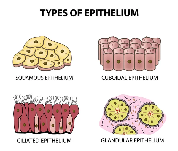
Types Of Epithelium. Squamous, Cubic, Ciliated, Glandular. Set. Infographics. Vector Illustration On Isolated Background
Vector, 2.27MB, 5000 × 4253 eps

Pseudostratified Epithelium Is A Type Of Epithelium That, Though Comprising Only A Single Layer Of Cells.
Image, 11.24MB, 5840 × 3893 jpg

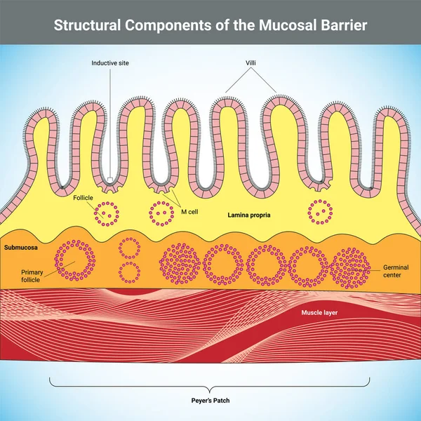
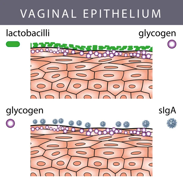
Mucosal Immune System Diagram. Mucous Or Gut Associated Lymphoid Tissue. Medical Vector Illustration
Vector, 3.14MB, 5000 × 5000 eps

3D Rendering Medically Accurate Illustration Of Intestinal Villi. Red Microvilli In A Intestinal Tract Organ Of The Gastrointestinal Banner. Isolated With Clipping Path.
Image, 2.97MB, 3500 × 2156 jpg
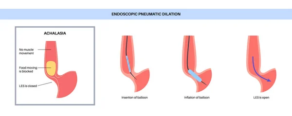
Endoscopic Pneumatic Dilation. Upper Endoscopy Minimally Invasive Procedure. Disorder Of The Esophagus, Therapy For Achalasia. Balloon Disrupts The Muscle Fibers In Closed Lower Esophageal Sphincter
Vector, 0.43MB, 6928 × 2768 eps
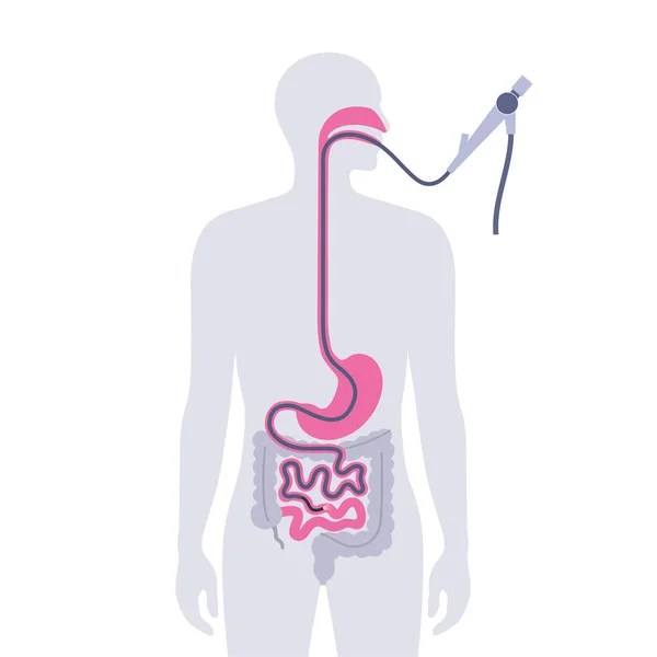
Esophagogastroduodenoscopy Medical Poster. Diagnostic Endoscopic Minimally Invasive Procedure. Visualization Of The Oropharynx, Esophagus, Stomach, And Proximal Duodenum. Gastroenterology Flat Vector
Vector, 0.41MB, 4168 × 4168 eps

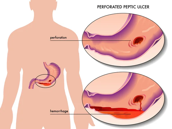
Five Stages Of The Development Of Oncological Disease - Cancer Of The Stomach
Vector, 4.64MB, 5683 × 5446 eps
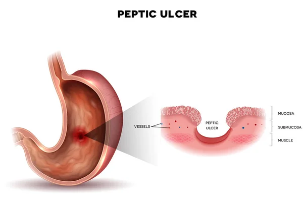

Progression Stages Of Stomach Cancer From Stage 0 To Stage IV, Highlighting Tumor Growth Diagram Hand Drawn Schematic Vector Illustration. Medical Science Educational Illustration
Vector, 0.72MB, 4000 × 4000 eps
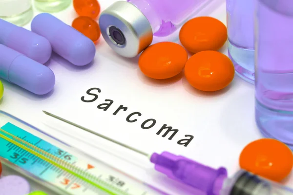
Sarcoma - Diagnosis Written On A White Piece Of Paper. Syringe And Vaccine With Drugs
Image, 8.85MB, 5184 × 3456 jpg
Page 1 >> Next