
Stock image Submucosa page 2

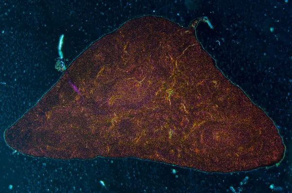
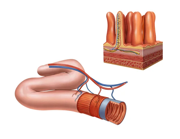
Duodenum Biopsy From The Pathology Of Small Intestine. Education For Human Histology.
Image, 13.92MB, 6000 × 4000 jpg


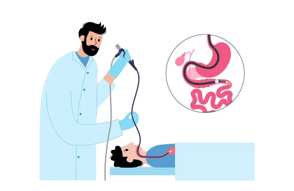
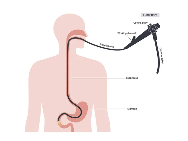
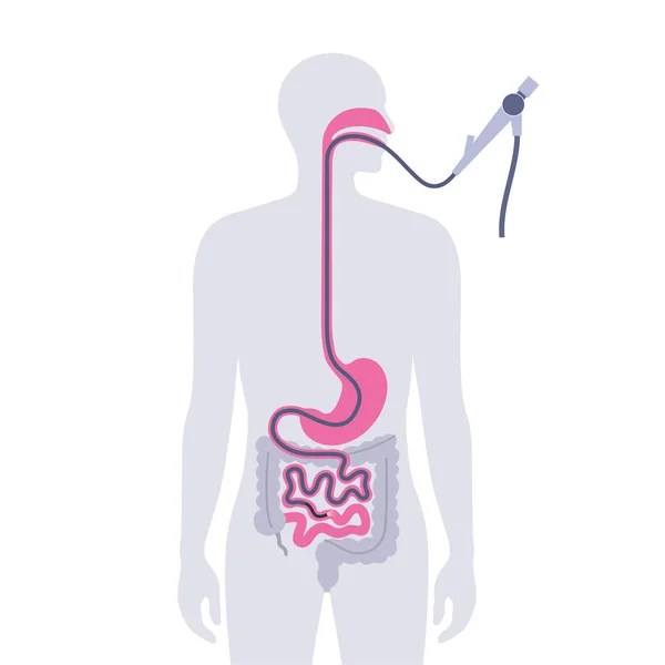
Esophagogastroduodenoscopy Medical Poster. Diagnostic Endoscopic Minimally Invasive Procedure. Visualization Of The Oropharynx, Esophagus, Stomach, And Proximal Duodenum. Gastroenterology Flat Vector
Vector, 0.64MB, 5840 × 3615 eps
Doctor's Hands In Blue Gloves Shows The Word Sarcoma. Medical Concept.
Image, 2.84MB, 3393 × 1986 jpg
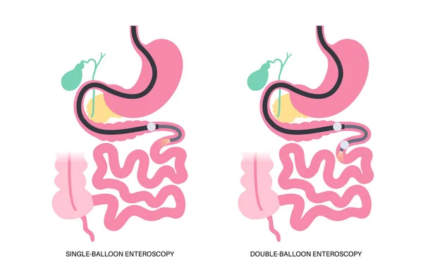
Double And Single Balloon Enteroscopy Minimally Invasive Procedure. Visualization Of The Small Intestine. Biopsy, Polyp Removal, Bleeding Therapy Or Stent Placement In Gastrointestinal Tract .poster.
Vector, 0.61MB, 5163 × 3365 eps
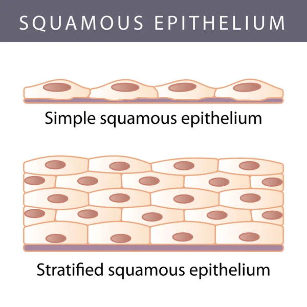
Epithelium. Squamous, Cubic, Ciliated, Glandular. Set. Infographics. Vector Illustration.
Vector, 1.56MB, 5000 × 5093 eps
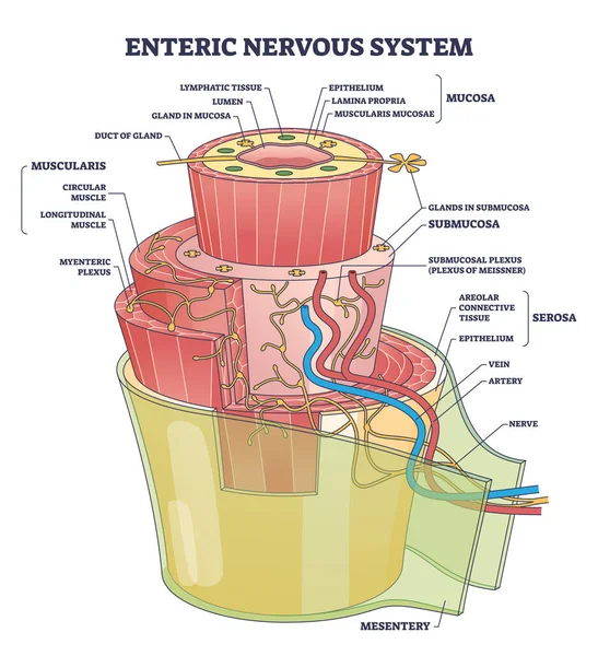
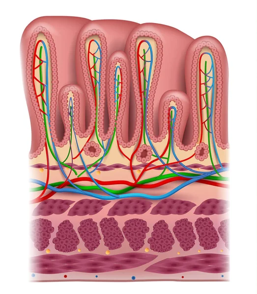
Enteric Nervous System Or ENS Intrinsic Autonomic Anatomy Outline Diagram. Labeled Educational Scheme With Complex Detailed Structure With Mucosa, Muscularis And Mesentery Part Vector Illustration.
Vector, 7.96MB, 4200 × 4600 eps

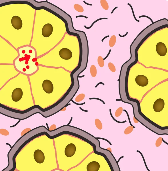
The Structure Of The Glandular Epithelium. Infographics. Vector Illustration On Isolated Background
Vector, 0.78MB, 5000 × 5000 eps
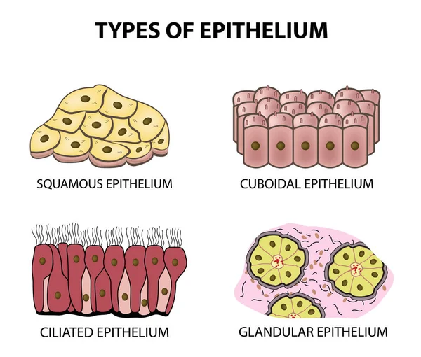
Types Of Epithelium. Squamous, Cubic, Ciliated, Glandular. Set. Infographics. Vector Illustration On Isolated Background
Vector, 2.27MB, 5000 × 4253 eps

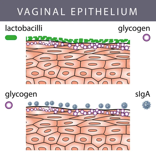
Mucosal Immune System Diagram. Mucous Or Gut Associated Lymphoid Tissue. Medical Vector Illustration
Vector, 3.14MB, 5000 × 5000 eps

3D Rendering Medically Accurate Illustration Of Intestinal Villi. Red Microvilli In A Intestinal Tract Organ Of The Gastrointestinal Banner. Isolated With Clipping Path.
Image, 2.97MB, 3500 × 2156 jpg
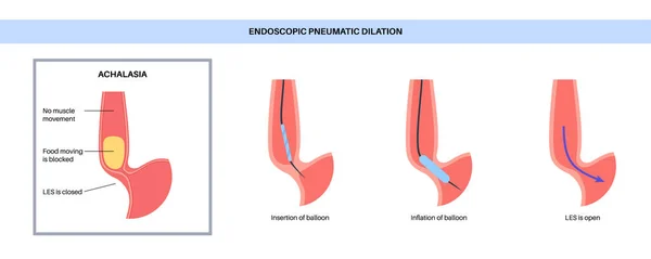
Endoscopic Pneumatic Dilation. Upper Endoscopy Minimally Invasive Procedure. Disorder Of The Esophagus, Therapy For Achalasia. Balloon Disrupts The Muscle Fibers In Closed Lower Esophageal Sphincter
Vector, 0.43MB, 6928 × 2768 eps

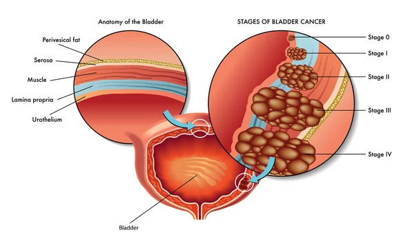
Five Stages Of The Development Of Oncological Disease - Cancer Of The Stomach
Vector, 4.64MB, 5683 × 5446 eps
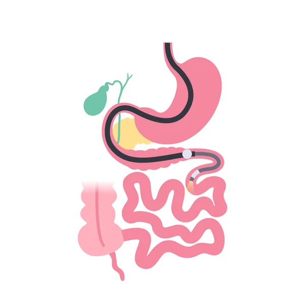
Balloon Assisted Enteroscopy. Visualization Of The Small Intestine Nonsurgical Procedure. Gastrointestinal Tract Exam. Biopsy, Polyp Removal, Bleeding Therapy Or Stent Placement Vector Illustration
Vector, 0.52MB, 5164 × 3365 eps
Esophagogastroduodenoscopy Medical Poster. Diagnostic Endoscopic Minimally Invasive Procedure. Visualization Of The Oropharynx, Esophagus, Stomach, And Proximal Duodenum. Gastroenterology Flat Vector
Vector, 0.46MB, 4826 × 3647 eps

Pseudostratified Epithelium Is A Type Of Epithelium That, Though Comprising Only A Single Layer Of Cells.
Image, 11.24MB, 5840 × 3893 jpg
Esophagogastroduodenoscopy Medical Poster. Diagnostic Endoscopic Minimally Invasive Procedure. Visualization Of The Oropharynx, Esophagus, Stomach, And Proximal Duodenum. Gastroenterology Flat Vector
Vector, 0.41MB, 4168 × 4168 eps
Balloon Assisted Enteroscopy. Visualization Of The Small Intestine Nonsurgical Procedure. Gastrointestinal Tract Exam. Biopsy, Polyp Removal, Bleeding Therapy Or Stent Placement Vector Illustration
Vector, 0.45MB, 4167 × 4167 eps
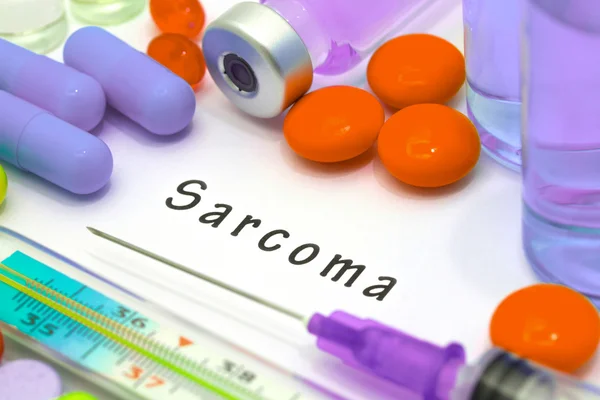
Sarcoma - Diagnosis Written On A White Piece Of Paper. Syringe And Vaccine With Drugs
Image, 8.85MB, 5184 × 3456 jpg

Esophagogastroduodenoscopy Medical Poster. Diagnostic Endoscopic Minimally Invasive Procedure. Visualization Of The Oropharynx, Esophagus, Stomach, And Proximal Duodenum. Gastroenterology Flat Vector
Vector, 0.47MB, 5164 × 3365 eps


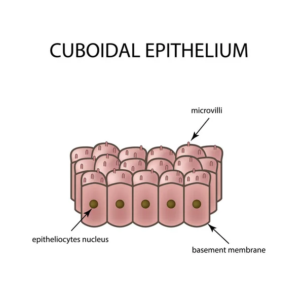
The Structure Of Cubic Epithelium. Infographics. Vector Illustration On Isolated Background
Vector, 1.62MB, 5000 × 5000 eps
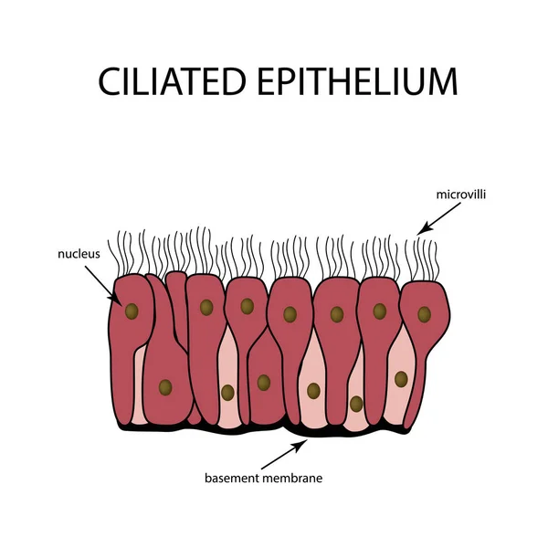
The Structure Of The Ciliated Epithelium. Infographics. Vector Illustration On Isolated Background
Vector, 0.49MB, 5000 × 5000 eps

Progression Stages Of Stomach Cancer From Stage 0 To Stage IV, Highlighting Tumor Growth Diagram Hand Drawn Schematic Raster Illustration. Medical Science Educational Illustration
Image, 4.19MB, 6000 × 6000 jpg
Previous << Page 2 >> Next