
Stock image Synthase
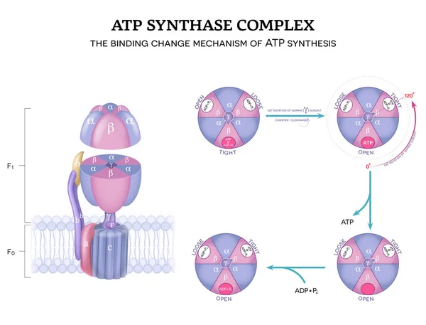
ATP Synthase Complex Structure And Mechanism Of ATP Synthase. The Binding Change Mechanism. 120-degree Rotation Of Gamma Subunit Counter-clockwise.
Vector, 9.35MB, 5512 × 4202 eps
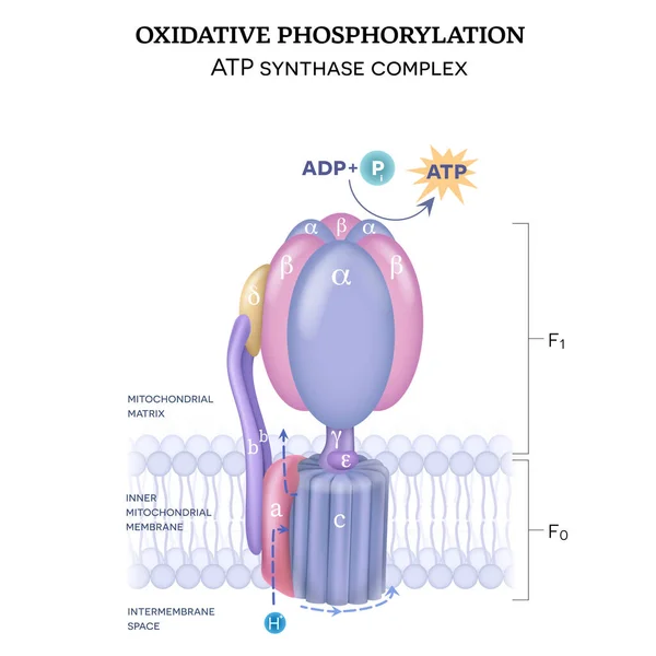
The ATP Synthase Structure (complex V) Consists Of Two Components F0 And F1. The Formation Of ATP Using Adenosine Diphosphate (ADP) And Inorganic Phosphate (Pi)
Vector, 11.37MB, 5000 × 5000 eps
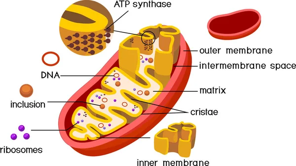
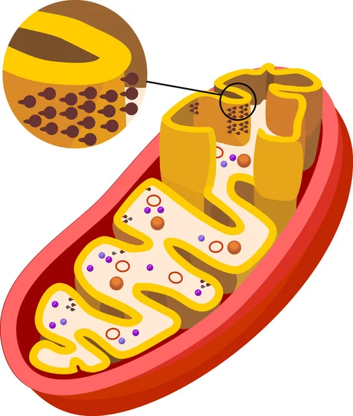
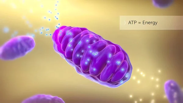
Structure Of Mitochondrion With ATP Synthase On Inner Membrane And Components Isolated On White Background
Vector, 5.74MB, 7580 × 4272 eps
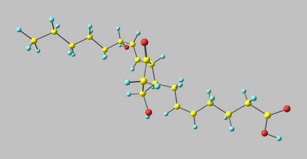
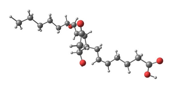
Prostaglandin D2 Or PGD2 Is A Prostaglandin That Binds To The Receptor PTGDR As Well As CRTH2. 3d Illustration
Image, 0.61MB, 7000 × 3638 jpg

Leucosceptrine Skeletal Structure Diagram.Sesterterpene Compound Molecule Scientific Illustration On White Background.
Vector, 5.4MB, 2000 × 2000 eps
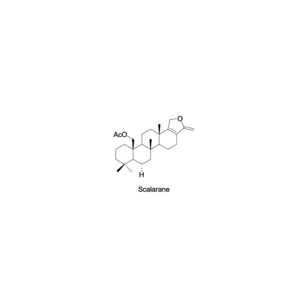
Scalarane Skeletal Structure Diagram.Sesterterpene Compound Molecule Scientific Illustration On White Background.
Vector, 5.39MB, 2000 × 2000 eps

Ophiobolin K Skeletal Structure Diagram.Sesterterpene Compound Molecule Scientific Illustration On Blue Background.
Vector, 6.35MB, 2000 × 2000 eps

Diagram Showing Enzymatic Transformation Of Steroid Hormones - Oestradiol To Oestrone And Oestrone Sulpfate. Biochemical Metabolic Endogenous Reaction.
Vector, 5.37MB, 2000 × 2000 eps

Diagram Showing Enzymatic Transformation Of Steroid Hormones - Oestradiol To Oestrone And Oestrone Sulpfate. Biochemical Metabolic Endogenous Reaction.
Vector, 5.65MB, 2000 × 2000 eps

Diagram Showing Enzymatic Transformation Of Steroid Hormones - Oestradiol To Oestrone And Oestrone Sulpfate. Biochemical Metabolic Endogenous Reaction.
Vector, 5.38MB, 2000 × 2000 eps

Diagram Showing Enzymatic Transformation Of Steroid Hormones - Oestradiol To Oestrone And Oestrone Sulpfate. Biochemical Metabolic Endogenous Reaction.
Vector, 5.66MB, 2000 × 2000 eps

Diagram Showing Enzymatic Transformation Of Steroid Hormones - Oestradiol To Oestrone And Oestrone Sulpfate. Biochemical Metabolic Endogenous Reaction.
Vector, 5.65MB, 2000 × 2000 eps
Prostaglandin D2 Or PGD2 Is A Prostaglandin That Binds To The Receptor PTGDR As Well As CRTH2. 3d Illustration
Image, 0.63MB, 7000 × 3638 jpg

Casbene Skeletal Structure Diagram.Sesquiterpene Compound Molecule Scientific Illustration On Yellow Background.
Vector, 5.37MB, 2000 × 2000 eps

Thorectandrol A Skeletal Structure Diagram.Sesterterpene Compound Molecule Scientific Illustration On Yellow Background.
Vector, 5.38MB, 2000 × 2000 eps

Diagram Showing Enzymatic Transformation Of Steroid Hormones - Oestradiol To Oestrone And Oestrone Sulpfate. Biochemical Metabolic Endogenous Reaction.
Vector, 5.65MB, 2000 × 2000 eps

Diagram Showing Enzymatic Transformation Of Steroid Hormones - Oestradiol To Oestrone And Oestrone Sulpfate. Biochemical Metabolic Endogenous Reaction.
Vector, 5.4MB, 2000 × 2000 eps

Sesterstatin 7 Skeletal Structure Diagram.Sesterterpene Compound Molecule Scientific Illustration On White Background.
Vector, 5.4MB, 2000 × 2000 eps

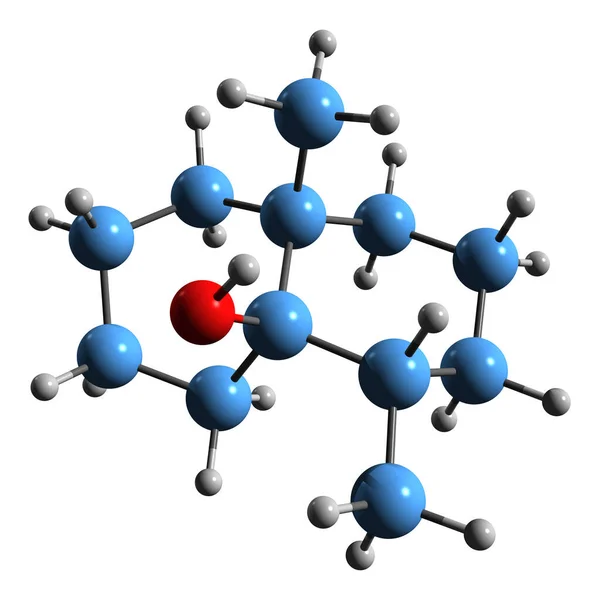
3D Image Of Geosmin Skeletal Formula - Molecular Chemical Structure Of Natural Bicyclic Terpene Isolated On White Background
Image, 4.34MB, 6452 × 6348 jpg
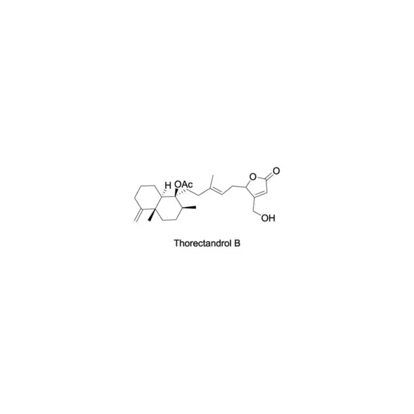
Thorectandrol B Skeletal Structure Diagram.Sesterterpene Compound Molecule Scientific Illustration On White Background.
Vector, 5.4MB, 2000 × 2000 eps

Detailed Citric Acid Cycle Pathway: Vector Illustration For Biochemistry, Molecular Biology, Health Science Education On White Background
Vector, 0.4MB, 5000 × 3000 ai

Cybastacines B Skeletal Structure Diagram.Sesterterpene Compound Molecule Scientific Illustration On Yellow Background.
Vector, 5.39MB, 2000 × 2000 eps

Steviol Skeletal Structure Diagram.Sesquiterpene Compound Molecule Scientific Illustration On Yellow Background.
Vector, 5.37MB, 2000 × 2000 eps

Diagram Showing Enzymatic Transformation Of Steroid Hormones - Oestradiol To Oestrone And Oestrone Sulpfate. Biochemical Metabolic Endogenous Reaction.
Vector, 5.66MB, 2000 × 2000 eps

Diagram Showing Enzymatic Transformation Of Steroid Hormones - Oestradiol To Oestrone And Oestrone Sulpfate. Biochemical Metabolic Endogenous Reaction.
Vector, 5.38MB, 2000 × 2000 eps

Diagram Showing Enzymatic Transformation Of Steroid Hormones - Oestradiol To Oestrone And Oestrone Sulpfate. Biochemical Metabolic Endogenous Reaction.
Vector, 5.38MB, 2000 × 2000 eps

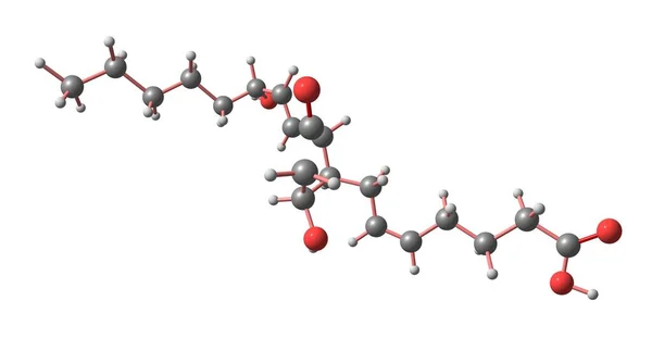
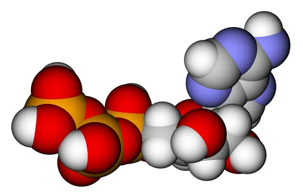
3D Image Of 5-Diphosphomevalonic Acid Skeletal Formula - Molecular Chemical Structure Of Intermediate In The Mevalonate Pathway Isolated On White Background
Image, 1.32MB, 5000 × 2451 jpg
Prostaglandin D2 Or PGD2 Is A Prostaglandin That Binds To The Receptor PTGDR As Well As CRTH2. 3d Illustration
Image, 0.65MB, 7000 × 3638 jpg
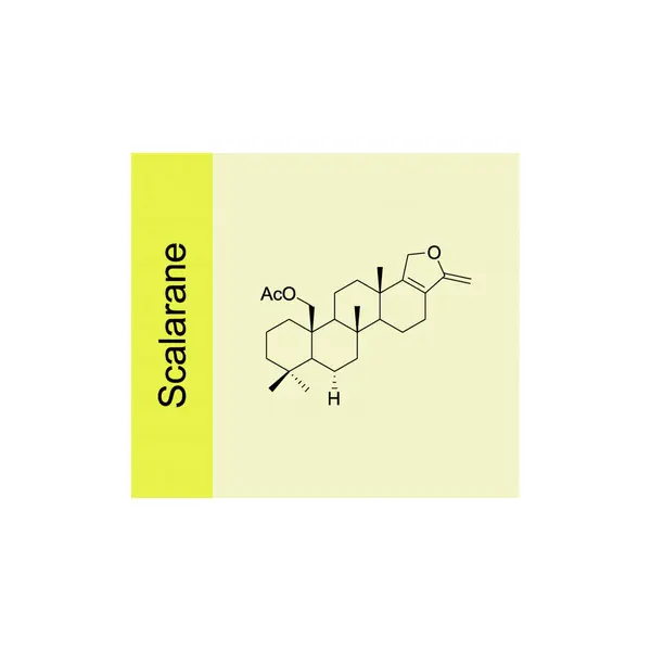
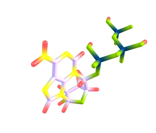
Scalarane Skeletal Structure Diagram.Sesterterpene Compound Molecule Scientific Illustration On Yellow Background.
Vector, 5.38MB, 2000 × 2000 eps

Eupheliotriol L Skeletal Structure Diagram.Diterpenoid Compound Molecule Scientific Illustration On Yellow Background.
Vector, 5.38MB, 2000 × 2000 eps

Diagram Showing Enzymatic Transformation Of Steroid Hormones - Oestradiol To Oestrone And Oestrone Sulpfate. Biochemical Metabolic Endogenous Reaction.
Vector, 5.39MB, 2000 × 2000 eps

Cybastacines A Skeletal Structure Diagram.Sesterterpene Compound Molecule Scientific Illustration On Blue Background.
Vector, 6.36MB, 2000 × 2000 eps

Scalarane Skeletal Structure Diagram.Sesterterpene Compound Molecule Scientific Illustration On Blue Background.
Vector, 6.01MB, 2000 × 2000 eps

Prostaglandin D2 Or PGD2 Is A Prostaglandin That Binds To The Receptor PTGDR As Well As CRTH2. 3d Illustration
Image, 0.62MB, 7000 × 3638 jpg

Biochemical Term Cofactors Written In White Bold Letters On Blue Background. 3d Illustration.
Image, 3.51MB, 3176 × 2000 jpg
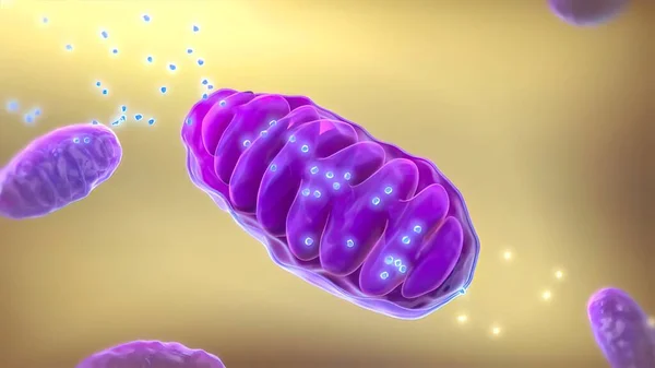
Structure Of Mitochondrion With ATP Synthase On Inner Membrane Isolated On White Background
Vector, 5.59MB, 5306 × 6240 eps

Ophiobolin K Skeletal Structure Diagram.Sesterterpene Compound Molecule Scientific Illustration On White Background.
Vector, 5.39MB, 2000 × 2000 eps

Diagram Showing Enzymatic Transformation Of Steroid Hormones - Oestradiol To Oestrone And Oestrone Sulpfate. Biochemical Metabolic Endogenous Reaction.
Vector, 5.37MB, 2000 × 2000 eps

Sesterstatin 7 Skeletal Structure Diagram.Sesterterpene Compound Molecule Scientific Illustration On Yellow Background.
Vector, 5.39MB, 2000 × 2000 eps

Leucosceptrine Skeletal Structure Diagram.Sesterterpene Compound Molecule Scientific Illustration On Yellow Background.
Vector, 5.4MB, 2000 × 2000 eps

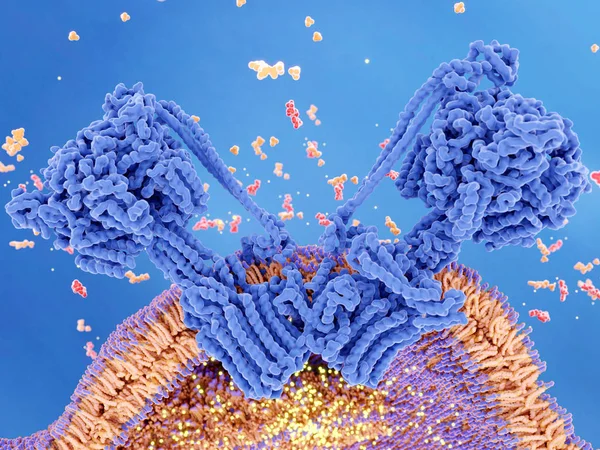
ATP Synthase Couples ATP (red) Synthesis From ADP And Inorganic Phosphate (orange) To A Proton Gradient (yellow) Created Across The Mitochondrial Membrane During Cellular Respiration.
Image, 12.41MB, 8000 × 6000 jpg

Thorectandrol B Skeletal Structure Diagram.Sesterterpene Compound Molecule Scientific Illustration On Yellow Background.
Vector, 5.39MB, 2000 × 2000 eps

Heliocide H Skeletal Structure Diagram.Sesterterpene Compound Molecule Scientific Illustration On White Background.
Vector, 5.39MB, 2000 × 2000 eps
Page 1 >> Next