
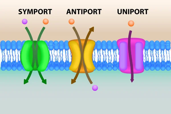
Stock image Transmembrane
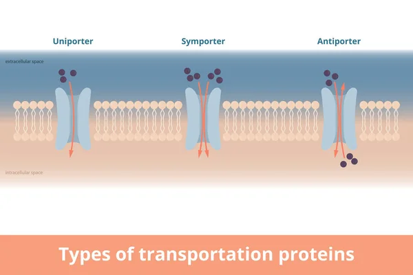
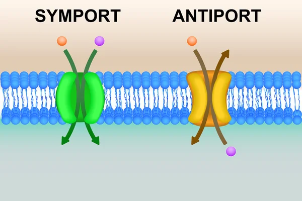
Types Of Cell Membrane Transportation Proteins. Visualization Of Uniporter (one Molecule, One Direction), Symporter (two Molecules, Same Directions), Antiporter (two Molecules, Different Directions).
Vector, 7.12MB, 6250 × 4167 eps

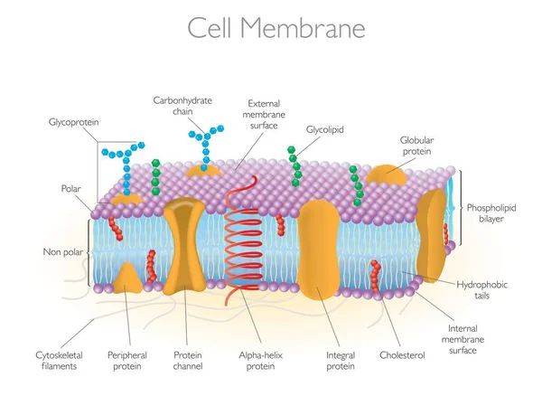
Membrane Proteins. Integral, And Peripheral Membrane Proteins, Single-pass, And Multi-pass Transmembrane Helix, Lipid-anchored Protein. Vector Illustration For Biological, Science And Educational Use
Vector, 4.52MB, 3921 × 3921 eps
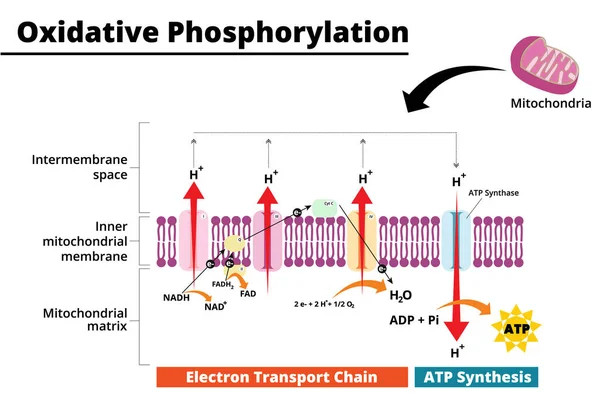
Oxidative Phosphorylation Process. Electron Transport Chain. The Final Step In Cellular Respiration. Vector Illustration. Didatic Illustration.
Vector, 0.73MB, 5000 × 3500 ai

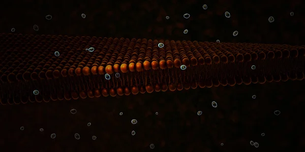
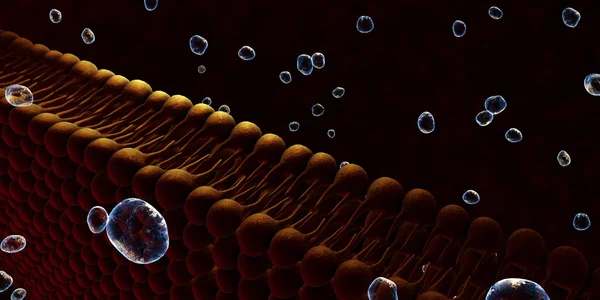
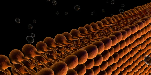
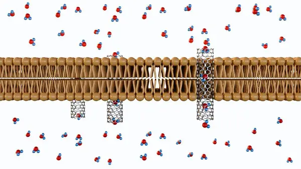
3d Rendering Of Molecules Passing Through Carbon Nanotube Porins On Lipid Bilayer Membrane
Image, 0.81MB, 3840 × 2160 jpg

3d Rendering Of Molecules Passing Through Carbon Nanotube Porins On Lipid Bilayer Membrane
Image, 0.57MB, 3840 × 2160 jpg
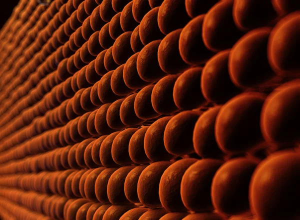
3d Rendering Of Gram Positive Bacteria Have A Thick Peptidoglycan Layer And No Outer Lipid Membrane
Image, 0.45MB, 3840 × 2160 jpg

3d Rendering Of Lipid Monolayer Is A Type Of Cell Membrane In Which The Lipids Are Arranged In A Single Layer, Rather Than The Typical Bilayer. Several Archaea Have A Lipid Monolayer
Image, 0.73MB, 3840 × 2160 jpg
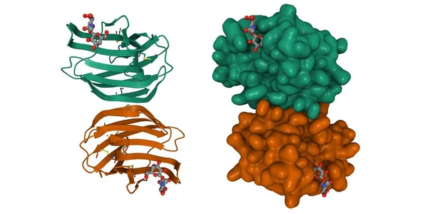
Crystal Structure Of Human Galectin-1 In Complex With Type 1 N-acetyllactosamine. 3D Cartoon And Gaussian Surface Model, Chain Id Color Scheme, PDB 4xbl, White Background
Image, 3.8MB, 8000 × 4000 jpg

3D Rendering Of Carbon Nanotube Porins, Short Pieces Of Carbon Nanotubes Capable Of Self-inserting Into A Lipid Bilayer, Model Of Biological Membrane Channels.
Image, 0.81MB, 3840 × 2160 jpg


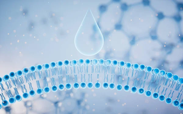
Cell Membrane With Blue Background, 3d Rendering. Computer Digital Drawing.
Image, 14.13MB, 8000 × 5000 jpg

3D Rendering Of Carbon Nanotube Porins, Short Pieces Of Carbon Nanotubes Capable Of Self-inserting Into A Lipid Bilayer, Model Of Biological Membrane Channels.
Image, 0.43MB, 3840 × 2160 jpg

3d Rendering Of Lipid Monolayer Is A Type Of Cell Membrane In Which The Lipids Are Arranged In A Single Layer, Rather Than The Typical Bilayer. Several Archaea Have A Lipid Monolayer
Image, 0.63MB, 3840 × 2160 jpg
Cell Membrane With Blue Background, 3d Rendering. Computer Digital Drawing.
Image, 13.09MB, 8000 × 5000 jpg

3D Rendering Of Carbon Nanotube Porins, Short Pieces Of Carbon Nanotubes Capable Of Self-inserting Into A Lipid Bilayer, Model Of Biological Membrane Channels.
Image, 0.69MB, 3840 × 2160 jpg
3d Rendering Of Lipid Monolayer Is A Type Of Cell Membrane In Which The Lipids Are Arranged In A Single Layer, Rather Than The Typical Bilayer. Several Archaea Have A Lipid Monolayer
Image, 0.77MB, 3840 × 2160 jpg

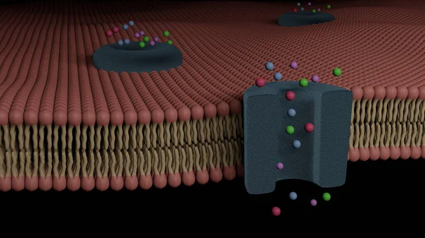
3d Rendering Of Molecules Passing Through Carbon Nanotube Porins On Lipid Bilayer Membrane
Image, 0.6MB, 3840 × 2160 jpg


3d Rendering Of Lipid Monolayer Is A Type Of Cell Membrane In Which The Lipids Are Arranged In A Single Layer, Rather Than The Typical Bilayer. Several Archaea Have A Lipid Monolayer
Image, 0.43MB, 3840 × 2160 jpg

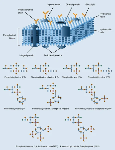
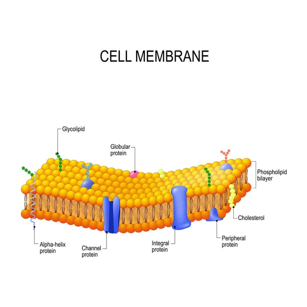
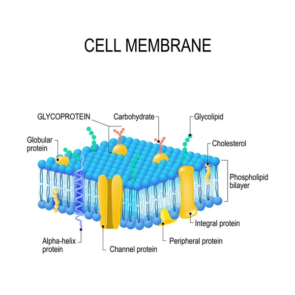
Detailed Structure Of Plasma Membrane: Vector Illustration For Biochemistry, Molecular Biology, Health Science Education On White Background
Vector, 0.74MB, 5000 × 3000 ai

The Atrial Muscle And Atrioventricular Node Belong To Fast And Slow Response Cells, Respectively, And Phase 0 Depolarization Is Responsible For Na+ Channels And Ca2+ Channels, Respectively.
Image, 6.25MB, 10000 × 6628 jpg
3d Rendering Of Molecules Passing Through Carbon Nanotube Porins On Lipid Bilayer Membrane
Image, 0.34MB, 3840 × 2160 jpg

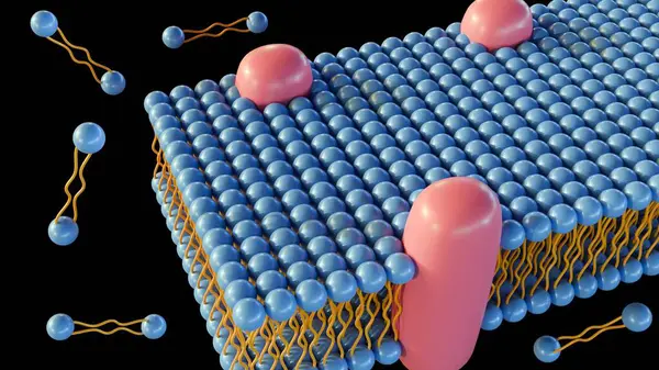
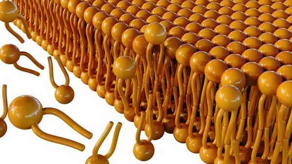
3d Rendering Of Assembly Or Formation Phospholipids Into Bilayer Structure
Image, 1.08MB, 3840 × 2160 jpg
3d Rendering Of Lipid Monolayer Is A Type Of Cell Membrane In Which The Lipids Are Arranged In A Single Layer, Rather Than The Typical Bilayer. Several Archaea Have A Lipid Monolayer
Image, 0.52MB, 3840 × 2160 jpg


3D Rendering Of Carbon Nanotube Porins, Short Pieces Of Carbon Nanotubes Capable Of Self-inserting Into A Lipid Bilayer, Model Of Biological Membrane Channels.
Image, 0.79MB, 3840 × 2160 jpg

3D Rendering Of Carbon Nanotube Porins, Short Pieces Of Carbon Nanotubes Capable Of Self-inserting Into A Lipid Bilayer, Model Of Biological Membrane Channels.
Image, 0.95MB, 3840 × 2160 jpg

3d Rendering Of Molecules Passing Through Carbon Nanotube Porins On Lipid Bilayer Membrane
Image, 0.67MB, 3840 × 2160 jpg
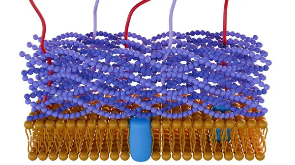
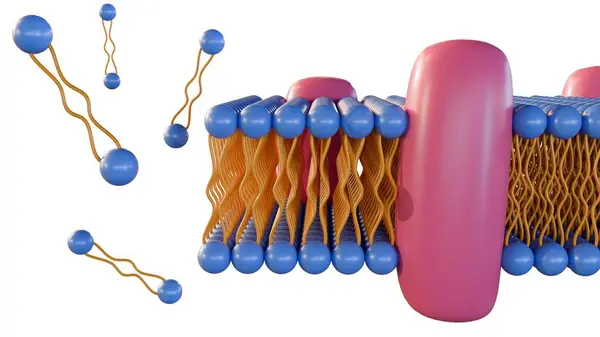
Glycoprotein Bilayer Cell Membrane Cross Section And Its Ion Channel (3D Rendering)
Image, 18.38MB, 8000 × 4500 jpg
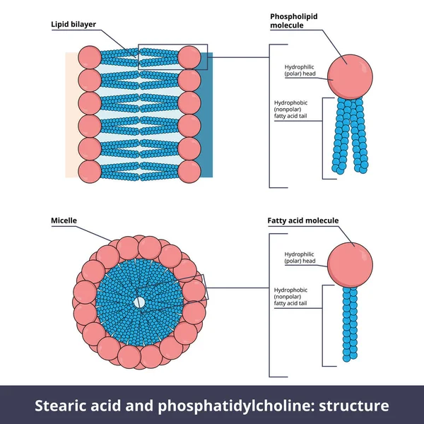
Structure Of Two Lipids. Stearic Acid (fatty Acid) And Phosphatidylcholine (phospholipid) Are Composed Of Chemical Groups That Form Polar Heads (hydrophilic) And Nonpolar Tails" (hydrophobic).
Vector, 10.84MB, 5208 × 5208 eps

3d Rendering Of Lipid Monolayer Is A Type Of Cell Membrane In Which The Lipids Are Arranged In A Single Layer, Rather Than The Typical Bilayer. Several Archaea Have A Lipid Monolayer
Image, 0.54MB, 3840 × 2160 jpg
3d Rendering Of Assembly Or Formation Phospholipids Into Bilayer Structure
Image, 1.66MB, 3840 × 2160 jpg
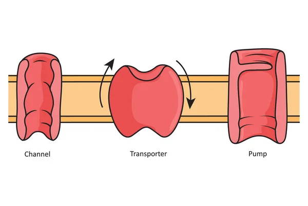
Channels, Transporters And Pumps, Simple Illustration Showing Different Transmembrane Proteins.
Image, 0.25MB, 3025 × 2025 jpg
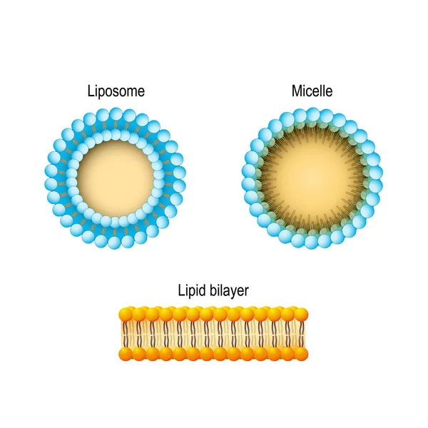
Cell Membrane (Lipid Bilayer), Micelle, Liposome. Phospholipids Aqueous Solution Structures. A Detailed Diagram Models Of Membrane Structure. Vector Illustration For Biology, Scientific, And Medical Use.
Vector, 8.35MB, 3969 × 3969 eps

Vector Illustration Of An Example Of Active Transport In Animal Cells - Sodium Potassium Pump.
Vector, 24.69MB, 3000 × 2254 eps

Passive Vs Active Cell Transport. Vector Illustration. Didatic Illustration.
Vector, 0.66MB, 5500 × 4500 ai

3D Rendering Of Carbon Nanotube Porins, Short Pieces Of Carbon Nanotubes Capable Of Self-inserting Into A Lipid Bilayer, Model Of Biological Membrane Channels.
Image, 0.4MB, 3840 × 2160 jpg

3d Rendering Of Molecules Passing Through Carbon Nanotube Porins On Lipid Bilayer Membrane
Image, 0.8MB, 3840 × 2160 jpg

3d Rendering Of Gram Positive Bacteria Have A Thick Peptidoglycan Layer And No Outer Lipid Membrane
Image, 0.54MB, 3840 × 2160 jpg
Page 1 >> Next