Home
Images
Vectors
Videos
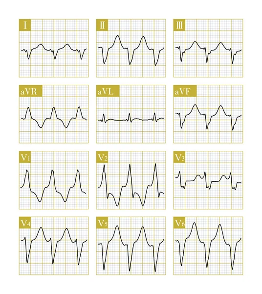
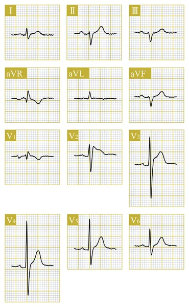
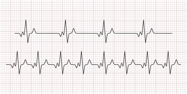
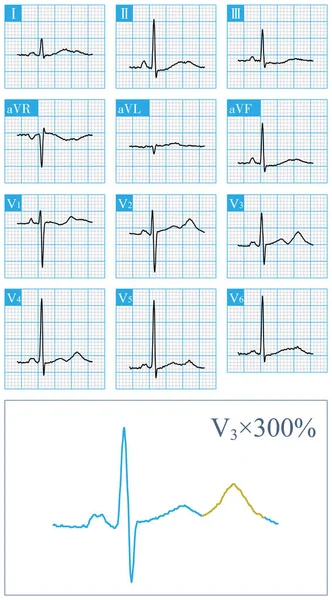
Stock image A 36 year old man survived CPR after sudden syncope. The electrocardiogram was suggestive of Brugada syndrome type 1. Implantation of ICD therapy.
Published:
Sep.19, 2022 06:47:34
Author:
asia11m
Views:
24
Downloads:
1
File type:
image
/
jpg
File size:
19.26 MB
Orginal size:
9000
x
13286
px
Available sizes:
S
(339 x 500)
M
(677 x 1000)
L
(1355 x 2000)
XL
(9000 x 13286)
EL0
(9000 x 13286)
Level:
beginner
Download Image
Image topics:
Human
Medicine
Medical
Cardiology
Heart
Monitor
Treatment
Death
Rhythm
Heartbeat
Cardiac
Ekg
Electrocardiogram
Ecg
Arrhythmia
Electrocardiograph
Cpr
Thrombosis
Myocardium
Heart Rate
Heart Disease
Cardiopulmonary Resuscitation
Syncope
Cardiac Arrest
Emergency Department
Sudden Death
Ventricular Fibrillation
Action Potential
Limb Leads
ECG Monitoring
Sodium Channel
Brugada Syndrome
Cardiovascular Medicine
St Segment
Ecg Education
Qrs Wave
T Wave Inversion
Malignant Ventricular Arrhythmia
St Segment Elevation
Cardiac Ion Channel Disease
Ion Channel Disease
Sodium Channel Gene Mutation
Scn5a Gene Mutation
Hereditary Heart Disease
Brugada Phenotype Ecg
Brudaga Syndrome
Ventricular Action Potential
J Point Elevation
Similar stock images
This Is A Case Of Ventricular Tachycardia In A Patient With Dilated Cardiomyopathy. This Ventricular Tachycardia Originates At The Apex Of The Heart, Negative QRS Main Wave In Leads V5 And V6.
10000 × 11187
Male, 66 Years Old, Received Chemotherapy For Esophageal Cancer. Type II Brugada Phenotype Appearsed On ECG, Which Needs To Be Differentiated From Primary Brugada Syndrome.
8500 × 13755
An Electrocardiogram Of A Patient With Pulmonary Embolism Showed That The Right Ventricle Was Overloaded, And The QRS Complex In The Chest Lead Was Dominated By S Waves.
10000 × 5791
Cardiogram. The Result Of Research CU Is Torn In Half It Is Isolated On A White Background.
4004 × 2558
A Is Infantile Type, Short ST Segment, Fast Fusion With T Wave; B Is A Wide Basal T Wave; C Is Normal Type; D Is Late Onset Normal Type, ST Segment Is Prolonged And T Wave Morphology Is Normal.
6500 × 6500
Cardiogram. Heart Beat. Heartbeat Line. Electrocardiogram. Vector
6976 × 3500
Medical Background. ECG. 4 Types Of Toning.
4341 × 2850
Female, 54 Years Old, Had Recurrent Syncope For 2 Years. The Clinical Diagnosis Was Type 2 Congenital Long QT Syndrome. Her Blood Potassium Is Normal, Do Not Misdiagnose Bimodal T Wave As U Wave.
4000 × 7228