
Stock image Limb Leads
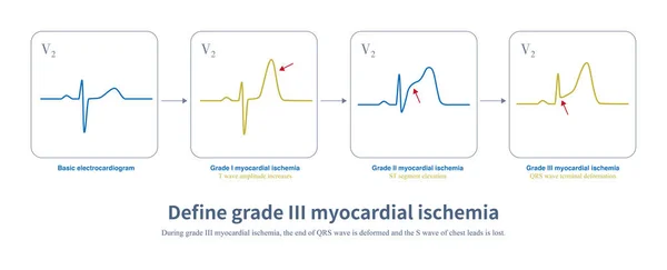
In Acute Myocardial Ischemia, The Amplitude Of T Wave Is Increased First, And Then The ST Segment Is Elevated. When The End Of QRS Wave Is Deformed, There Is A Lack Of Collateral Circulation.
Image, 1.32MB, 10108 × 4093 jpg

Electrocardiography Vector Concept. ECG Limb Leads Flat Illustration
Vector, 1.36MB, 5121 × 4000 eps

Ventricular Tachyarrhythmia Includes Many Clinical Types, Some Benign And Some Malignant. For Malignant Ventricular Arrhythmias, Patients Are At Risk Of Death.
Image, 27.66MB, 8000 × 10973 jpg

The QT Interval Of ECG Is From The Beginning Of QRS Wave To The End Of T Wave, Representing The Total Time Of Ventricular Depolarization And Repolarization.
Image, 8.09MB, 10000 × 10950 jpg

A 36 Year Old Man Survived CPR After Sudden Syncope. The Electrocardiogram Was Suggestive Of Brugada Syndrome Type 1. Implantation Of ICD Therapy.
Image, 19.26MB, 9000 × 13286 jpg

During Left Posterior Fascicular Block, The ECG Showed Right Axis Deviation. The QRS Wave In Leads I And AVL Was RS Wave, And The Duration Of QRS Wave Was Less Than 120 Ms.
Image, 30.53MB, 10000 × 11472 jpg

R Wave Greater Than S Wave Is Judged To Be Positive; R Smaller Than S Is Judged To Be Negative; R Equal To S Amplitude Is Judged To Be Equipotential.
Image, 7.5MB, 10000 × 5119 jpg
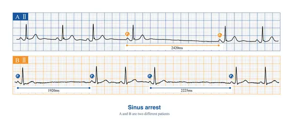
When Sinus Arrest Occurs, The Electrocardiogram Will Show A Long P-P Interval, Which Is Not Multiples Of The Basal Sinus Cycle, Including Physiological And Pathological Reasons.
Image, 8.96MB, 10000 × 4418 jpg
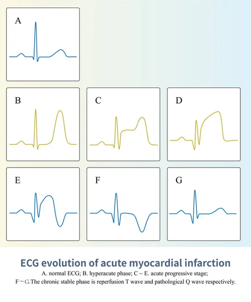
In ST Segment Elevation Myocardial Infarction, The ST-T Of ECG Will Undergo A Characteristic Evolution Process, And Finally Appear Pathological Q Wave, Sometimes Lasting For A Lifetime.
Image, 7.27MB, 8000 × 9619 jpg

When The Frontal QRS Axis Is At +83, The R Amplitude Of Lead AVF Is The Highest.The Frontal QRS Axis Is Almost Perpendicular To The Axis Of Lead .
Image, 11.75MB, 10000 × 6576 jpg
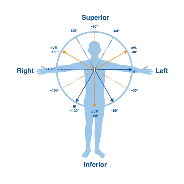
In The Frontal Lead System, The Lead Axes Of The 6 Limb Leads Form A Hexaxial Reference System, Which Is One Of The Important Theories Of Electrocardiography.
Image, 8.21MB, 10000 × 10000 jpg

When The Limb Leads Are Reversed, It Is Common For The Left Upper Limb And The Right Upper Limb To Be Reversed, Affecting The ECG Pattern Of The Limb Leads.
Image, 9.69MB, 10000 × 5625 jpg

Vector Illustration Of A Disabled Girl With A Prosthetic Leg Leads An Active Lifestyle And Goes Hiking In The Forest. Rehabilitation And Adaptation Of People With Disabilities.
Vector, 1.2MB, 5657 × 4000 eps

When The Limb Leads Are Reversed, It Is Common For The Left Upper Limb And The Right Upper Limb To Be Reversed, Affecting The ECG Pattern Of The Limb Leads.
Image, 12.91MB, 10000 × 11438 jpg
Page 1 >> Next