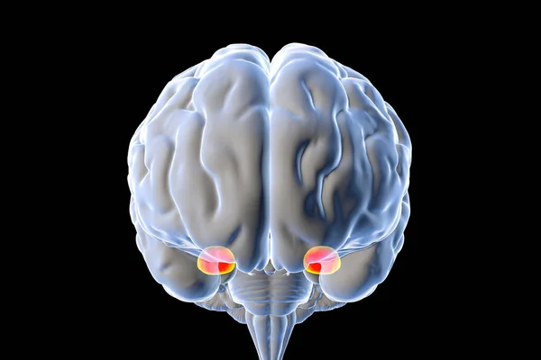
Stock image Amygdala, also known as corpus amygdaloideum, in the brain, 3D illustration. Two almond-shaped clusters of nuclei within the temporal lobes, part of the limbic system, play role in memory and emotions
Published: Jan.14, 2021 09:08:05
Author: katerynakon
Views: 299
Downloads: 21
File type: image / jpg
File size: 2.47 MB
Orginal size: 6000 x 4000 px
Available sizes:
Level: silver