photo free
Home Images Vectors Videos

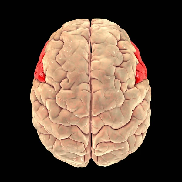
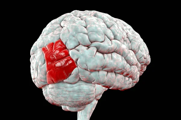
Stock image Human brain with highlighted superior occipital gyrus, 3D illustration. It is located in occipital lobe and is responsible for object recognition
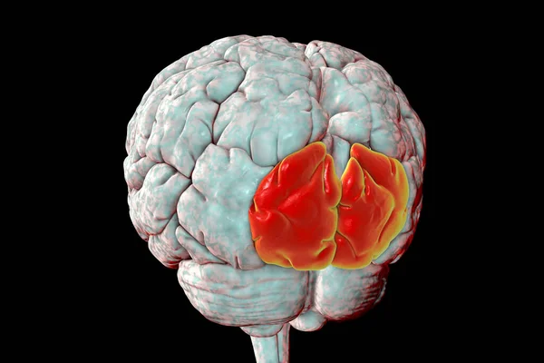
Stock image Human brain with highlighted superior occipital gyrus, 3D illustration. It is located in occipital lobe and is responsible for object recognition
Published: Dec.01, 2020 16:21:16
Author: katerynakon
Views: 25
Downloads: 1
File type: image / jpg
File size: 4.49 MB
Orginal size: 6000 x 4000 px
Available sizes:
S
(500 x 333)
M
(1000 x 667)
L
(2000 x 1333)
XL
(6000 x 4000)
EL0
(6000 x 4000)
Level: silver
Download Image
Image topics:

Similar stock images

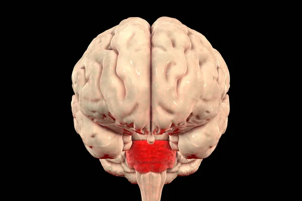
stock image Pons Varolii highlighted inside human brain, 3D illustration. It part of the brainstem, includes neural pathways carrying signals from the brain down to cerebellum and medulla, and into the thalamus

Pons Varolii Highlighted Inside Human Brain, 3D Illustration. It Part Of The Brainstem, Includes Neural Pathways Carrying Signals From The Brain Down To Cerebellum And Medulla, And Into The Thalamus

6000 × 4000
stock image Human brain with highlighted superior parietal lobule, 3D illustration

Human Brain With Highlighted Superior Parietal Lobule, 3D Illustration

4400 × 4400
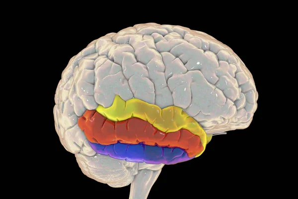
stock image Human brain with highlighted temporal gyri, 3D illustration. Superior temporal gyrus (yellow), middle (red), and inferior (blue). They are involved in processing auditory information and encoding of memory

Human Brain With Highlighted Temporal Gyri, 3D Illustration. Superior Temporal Gyrus (yellow), Middle (red), And Inferior (blue). They Are Involved In Processing Auditory Information And Encoding Of Memory

6000 × 4000
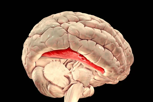
stock image Human brain with highlighted fusiform gyrus, or medial occipitotemporal gyrus, 3D illustration. It is associated with various neural pathways related to recognition, and also to synesthesia, dyslexia, and prosopagnosia

Human Brain With Highlighted Fusiform Gyrus, Or Medial Occipitotemporal Gyrus, 3D Illustration. It Is Associated With Various Neural Pathways Related To Recognition, And Also To Synesthesia, Dyslexia, And Prosopagnosia

6000 × 4000
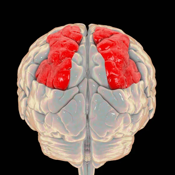
stock image Human brain with highlighted inferior frontal gyrus, top view, 3D illustration. It is a part of the prefrontal cortex and the location of Broca's area, involved in language processing and speech production

Human Brain With Highlighted Inferior Frontal Gyrus, Top View, 3D Illustration. It Is A Part Of The Prefrontal Cortex And The Location Of Broca's Area, Involved In Language Processing And Speech Production

5000 × 5000
stock image Human brain with highlighted middle (lateral) occipital gyrus, 3D illustration. It is located in occipital lobe and is responsible for object recognition

Human Brain With Highlighted Middle (lateral) Occipital Gyrus, 3D Illustration. It Is Located In Occipital Lobe And Is Responsible For Object Recognition

6202 × 4135
stock image Human brain with highlighted superior occipital gyrus, 3D illustration. It is located in occipital lobe and is responsible for object recognition

Human Brain With Highlighted Superior Occipital Gyrus, 3D Illustration. It Is Located In Occipital Lobe And Is Responsible For Object Recognition

6349 × 4233