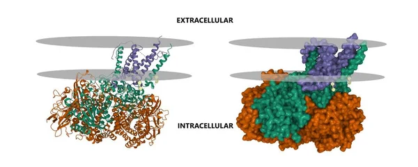
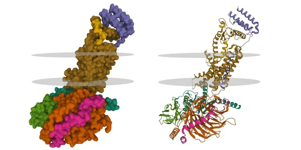
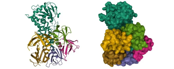
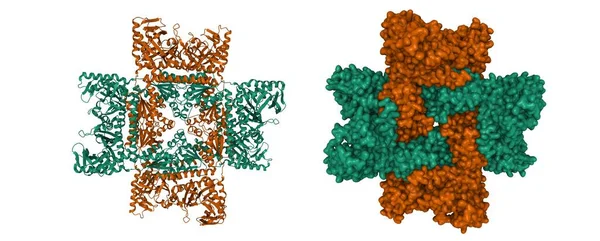
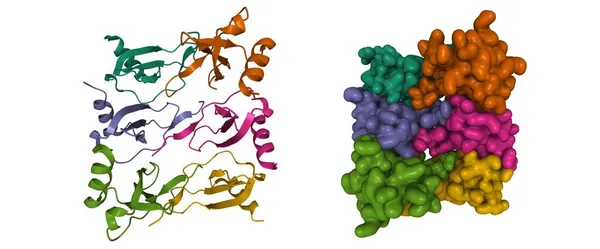
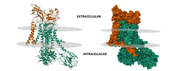
Stock image Cryo-EM structure of a human ATP11C(green)-CDC50A(brown) flippase in E1AlF state. 3D cartoon and Gaussian surface models, PDB 7bss, white background
Published: Oct.18, 2021 13:15:03
Author: unnaugan
Views: 2
Downloads: 0
File type: image / jpg
File size: 4.99 MB
Orginal size: 10000 x 4000 px
Available sizes:
Level: beginner