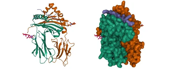
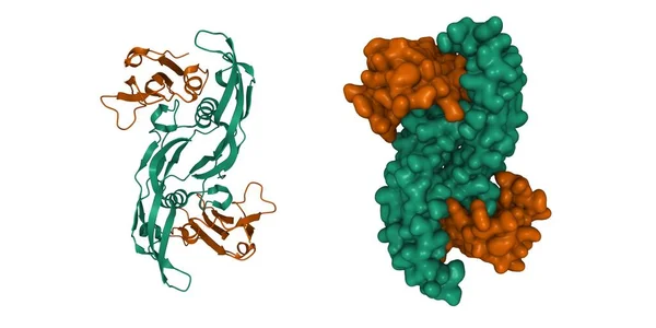
Stock image Crystal structure of BMP-2 (green) in complex with BMPR-IA variant B12 (brown). 3D cartoon and Gaussian surface models, PDB 2qja, white background
Published: Dec.21, 2021 12:14:53
Author: unnaugan
Views: 1
Downloads: 0
File type: image / jpg
File size: 3.42 MB
Orginal size: 8000 x 4000 px
Available sizes:
Level: beginner