Home
Images
Vectors
Videos
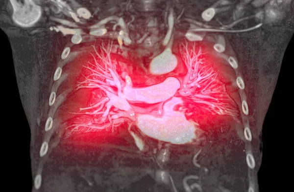
Stock image CT Chest or Lung 3d rendering image showing Trachea and lung in respiratory system.
Published:
Apr.27, 2024 04:06:02
Author:
samunella
Views:
0
Downloads:
0
File type:
image
/
jpg
File size:
1.65 MB
Orginal size:
4096
x
2160
px
Available sizes:
S
(500 x 264)
M
(1000 x 527)
L
(2000 x 1055)
XL
(4096 x 2160)
EL0
(4096 x 2160)
Level:
beginner
Download Image
Image topics:
Oxygenation
Blood Vessel
Respiratory System
Blood Flow
Circulatory System
Pulmonary Trunk
Pulmonary Veins
Right Ventricle
Arterial Blood
Oxygen Supply
Pulmonary Hypertension
Main Artery
Pulmonary Circulation
Pulmonary Artery
Pulmonary Disease
Heart Pumping
Oxygen Exchange
Cardiovascular Health
Cardiac Output
Deoxygenated Blood
Heart Function
Heart And Lungs
Vascular Anatomy
Pulmonary Valve
Cardiopulmonary System
Cardiac Function
Cardiovascular Physiology
Oxygenated Blood
Oxygen Delivery
Oxygen Rich Blood
Cardiovascular Anatomy
Vascular Health
Respiratory Exchange
Vascular Function
Lung Capillaries
Lung Connection
Arterial Pathway
Oxygenation Process
Oxygen Transport
Ventricular Connection
Vital Transport
Aorta Branch
Arterial Branching
Transport Of Gases
Vascular Pathway
Blood Oxygen Levels
Vital Pathway
Lung Arteries
Arterial Function
Similar stock images
A CTA Pulmonary Artery Reveals A Detailed View Of The Lung Blood Vessels, Capturing The Presence Of A Pulmonary Embolism, A Condition Where A Blood Clot Disrupts Normal Blood Flow.
4096 × 2160
CTPA Or CTA Pulmonary Artery .This Imaging Technique Offers A Clear View Of The Pulmonary Arteries, Aiding In The Diagnosis Of Pulmonary Embolism, Vascular Conditions, And Other Respiratory Issues.
3840 × 2160
CT Upper Abdomen Coronal View Showing DDX Is Atypical HCC Or Hepatocellular Carcinoma.
5594 × 3275
MRI Of The Upper Abdomen Coronal View Is A Non-invasive Imaging Technique Providing Detailed Visuals Of Organs Like The Liver, Pancreas, And Kidneys.
3840 × 2160
A CTA Pulmonary Artery Reveals A Detailed View Of The Lung Blood Vessels, Capturing The Presence Of A Pulmonary Embolism, A Condition Where A Blood Clot Disrupts Normal Blood Flow.
4096 × 2160
CT Upper Abdomen Coronal Showing DDX Is Atypical HCC Or Hepatocellular Carcinoma.
3080 × 2866
A CTA Pulmonary Artery Reveals A Detailed View Of The Lung Blood Vessels, Capturing The Presence Of A Pulmonary Embolism, A Condition Where A Blood Clot Disrupts Normal Blood Flow.
4096 × 2160
CTA Pulmonary Arteries 3D Rendering Showing Branch Of Pulmonary Artery
4707 × 3088