
Stock image Aorta Branch
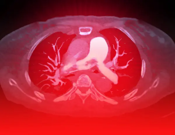
A CTA Pulmonary Artery Reveals A Detailed View Of The Lung Blood Vessels, Capturing The Presence Of A Pulmonary Embolism, A Condition Where A Blood Clot Disrupts Normal Blood Flow.
Image, 2.47MB, 3856 × 2976 jpg

A CTA Pulmonary Artery Reveals A Detailed View Of The Lung Blood Vessels, Capturing The Presence Of A Pulmonary Embolism, A Condition Where A Blood Clot Disrupts Normal Blood Flow.
Image, 3.49MB, 5352 × 3229 jpg

The Doctor Uses The Image As An Educational Tool, Simplifying The Concept Of Pulmonary Embolism To Ensure Patients Comprehend The Diagnosis.Clipping Path.
Image, 3.15MB, 3904 × 3117 jpg
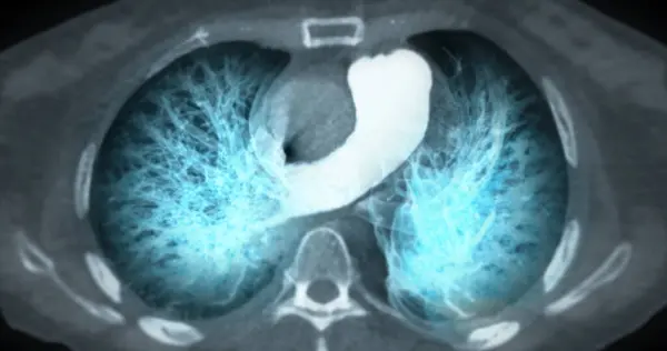
CT Chest Or Lung 3d Rendering Image Showing Trachea And Lung In Respiratory System.
Image, 1.53MB, 4096 × 2160 jpg

A CTA Pulmonary Artery Reveals A Detailed View Of The Lung Blood Vessels, Capturing The Presence Of A Pulmonary Embolism, A Condition Where A Blood Clot Disrupts Normal Blood Flow.
Image, 4.21MB, 3904 × 3117 jpg

The Hand In The Image Guides Your Attention To The Tablet, Where A Visual Representation Depicts Pulmonary Embolism, Aiding In Easy Understanding.Clipping Path.
Image, 1.98MB, 3904 × 3117 jpg

A CTA Pulmonary Artery Reveals A Detailed View Of The Lung Blood Vessels, Capturing The Presence Of A Pulmonary Embolism, A Condition Where A Blood Clot Disrupts Normal Blood Flow.
Image, 2.63MB, 4032 × 3024 jpg

CT Chest Or Lung 3d Rendering Image Showing Trachea And Lung In Respiratory System.
Image, 1.65MB, 4096 × 2160 jpg

A CTA Pulmonary Artery Reveals A Detailed View Of The Lung Blood Vessels, Capturing The Presence Of A Pulmonary Embolism, A Condition Where A Blood Clot Disrupts Normal Blood Flow.
Image, 2.64MB, 4096 × 2160 jpg

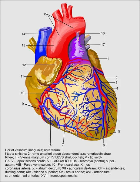


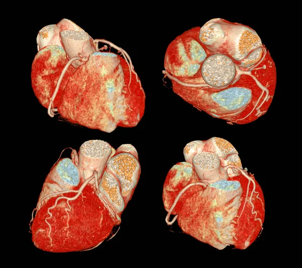
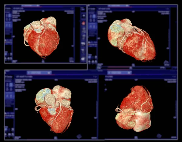
CT Cardiac 3D Or CTA Coronary Artery For Prevention And Screening Coronary Artery Diseases.
Image, 4.27MB, 3840 × 3408 jpg
CT Cardiac 3D Or CTA Coronary Artery For Prevention Coronary Artery Diseases.
Image, 2.33MB, 3840 × 3408 jpg
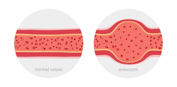
Healthy Vessel And Sick Vessel With Aneurysm With Blood Cells Flat Vector Illustration
Vector, 3.02MB, 6547 × 3208 eps
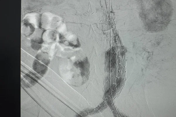
X-ray Image. Angiogram Of Right Common Iliac Artery After Aortic Stent Graft Deployed At Infra Renal Abdominal Aortic Aneurysm During EVAR
Image, 19.11MB, 6000 × 4000 jpg


Healthy Vessel And Sick Vessel With Aneurysm With Blood Cells Flat Vector Illustration
Vector, 3.03MB, 6547 × 3208 eps

X-ray Image. Angiogram Of Right Common Iliac Artery After Aortic Stent Graft Deployed At Infra Renal Abdominal Aortic Aneurysm During EVAR
Image, 20.71MB, 6000 × 4000 jpg


Cooking, Medicine, Education And Other Web Icon In Outline Style.glow, Travel, Finance Icons In Set Collection.
Vector, 4.82MB, 5000 × 5000 eps

Angiogram Of Right Common Iliac Artery After Aortic Stent Graft Deployed At Infra Renal Abdominal Aortic Aneurysm During EVAR Procedure.
Image, 14.57MB, 6000 × 4000 jpg

X-ray Image. Angiogram Of Right Common Iliac Artery After Aortic Stent Graft Deployed At Infra Renal Abdominal Aortic Aneurysm During EVAR
Image, 16.34MB, 5040 × 4000 jpg

Mesenteric Artery Stenosis As Blockage In Blood Vessel Outline Diagram. Labeled Educational Scheme With Dangerous Medical Condition For Abdomen And Digestive Tract Health Vector Illustration.
Vector, 6.48MB, 4400 × 4000 eps

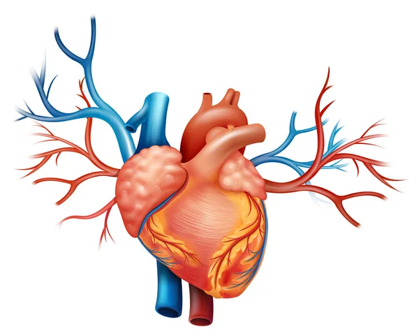
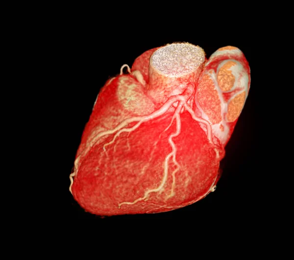
CTA Coronary Artery 3D Rendering Is A Diagnostic Imaging Technique Capturing Detailed Visuals Of The Heart's Blood Vessels In Diagnosing Coronary Artery Diseases And Assessing Cardiac Health.
Image, 1.74MB, 3840 × 2646 jpg


Mesenteric Artery Anatomy And Abdominal Aorta Location Outline Diagram. Labeled Educational Medical Scheme With Abdomen And Bowel Blood Flow Vector Illustration. Ileocolical And Jejunal Arteries.
Vector, 6.09MB, 4400 × 4000 eps

Male, 13 Years Old, Clinically Diagnosed With Secundum Atrial Septal Defect. Note That The QRS Wave In Lead V1 Of The Electrocardiogram Has A QR Shape, Indicating Right Ventricular Hypertrophy.
Image, 7.37MB, 10000 × 7203 jpg
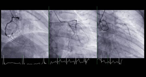
Cardiac Catheterization Can Help Doctor Diagnose And Treat Problems In Your Heart And Blood Vessels That Might Otherwise Cause Larger Issues, Such As A Heart Attack Or Stroke.
Image, 1.62MB, 4096 × 2160 jpg

Page 1 >> Next